Cleavage
Cytoplasmic Anomalies
Cytoplasmic Anomalies
-
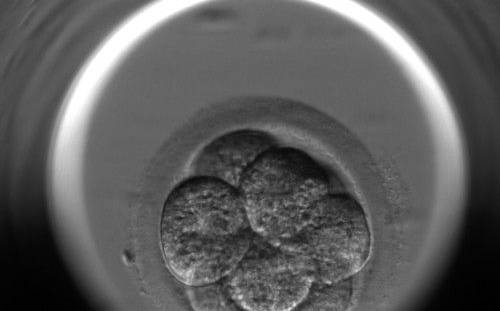
Figure 271
An 8-cell embryo with equally sized blastomeres showing cytoplasmic pitting. Numerous small pits are present on the surface of the cytoplasm.
-
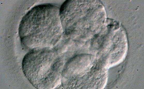
Figure 272
An 8-cell embryo with equally sized blastomeres showing cytoplasmic pitting. Numerous small pits are homogeneously distributed in the cytoplasm. The 5 blastomeres in focus are arranged in one spatial plane.
-
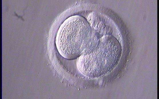
Figure 273
A 2-cell embryo with a clear halo in both blastomeres, characterized by centralized granularity associated with an absence of organelles in the peripheral cortex.
-
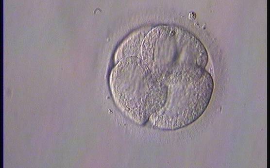
Figure 274
A 4-cell embryo on Day 2 with an abnormal distribution of organelles leading to differential granular and smooth zones inside each cell.
-
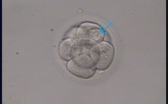
Figure 275
An 8-cell embryo with one blastomere showing a small vacuole (arrow).
-
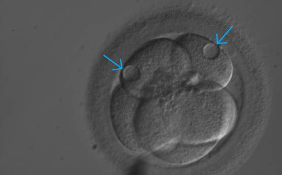
Figure 276
A 5-cell embryo with two small and three large blastomeres. There is a small vacuole in each of the two smaller blastomeres.
-
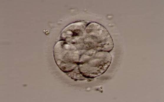
Figure 277
An embryo with abundant small vacuoles.
-
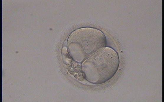
Figure 278
A 2-cell embryo with large vacuoles in both blastomeres and 15% concentrated fragmentation.
-
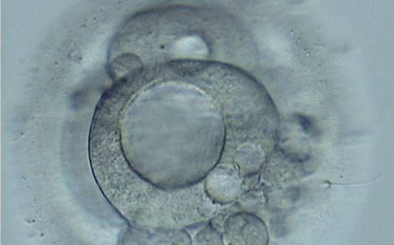
Figure 279
A 3-cell embryo with a large vacuole in the blastomere in the first plane in this view. A smaller vacuole is present in another blastomere. High fragmentation, about 40%, concentrated in one area.
-
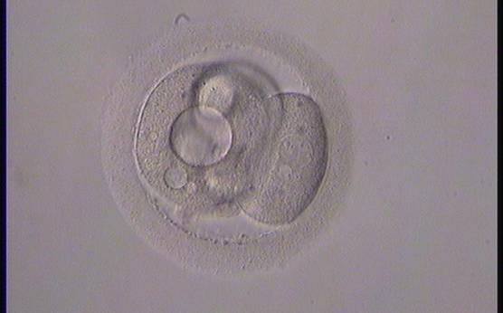
Figure 280
A 3-cell embryo with different sized blastomeress showing both large and small vacuoles.