Cleavage
Compaction
Compaction
-
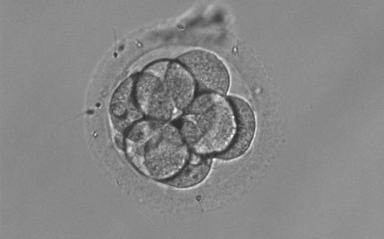
Figure 291
An 8-cell embryo that shows no signs of compaction. Cells are evenly sized and barely touching.
-
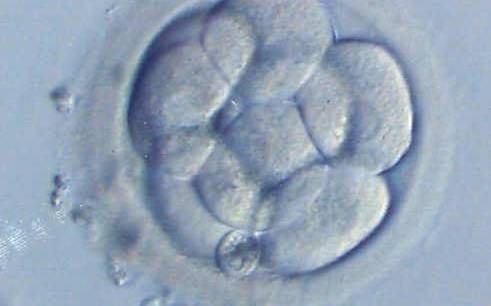
Figure 292
An 8-cell embryo showing signs of initial compaction. The cells are tightening their contact.
-
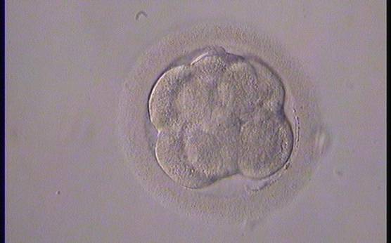
Figure 293
An 8-cell embryo showing signs of moderate compaction. Individual blastomeres are becoming difficult to identify.
-
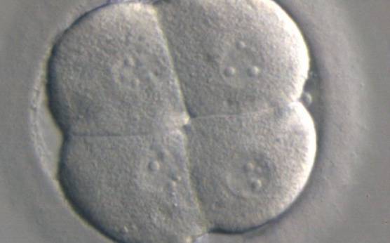
Figure 294
A clover shaped 4-cell embryo showing signs of very early compaction. A single nucleus is clearly visible in each cell.
-
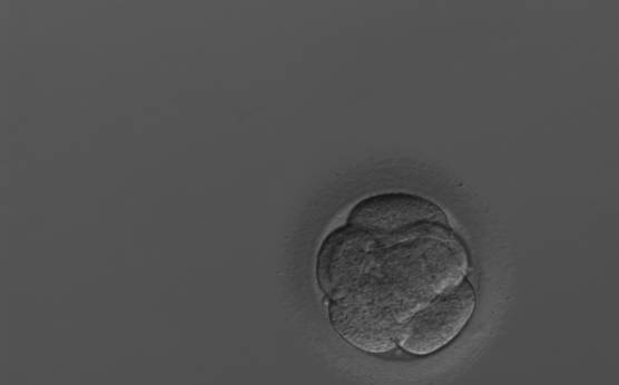
Figure 295
A 7-cell embryo showing signs of early compaction. This embryo was transferred but failed to implant.
-
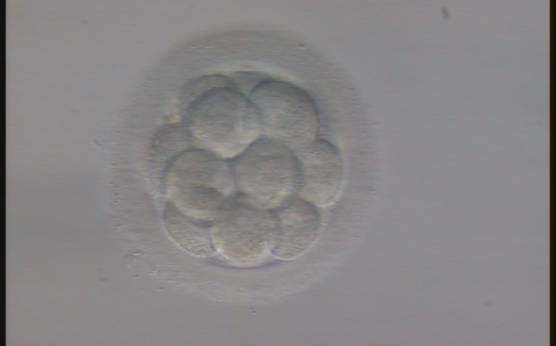
Figure 296
An embryo with more than 12 blastomeres showing no signs of compaction. With this number of cells it is very unusual that the embryo has not yet initiated compaction.
-
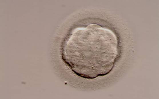
Figure 297
A morula of good quality. All blastomeres have been included in the compaction process and individual cells are no longer evident.
-
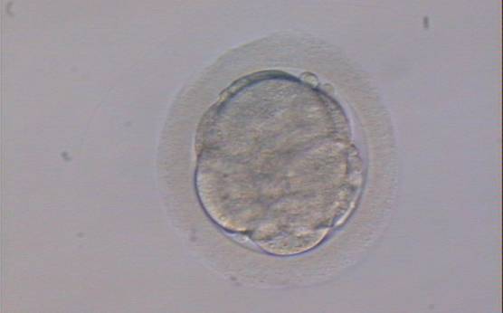
Figure 298
A fair quality morula. Some cell boundaries are still visible and a few small cells (or fragments) are not completely incorporated into the compaction process.
-
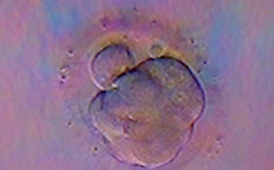
Figure 299
A fair quality morula. Cell boundaries are still visible and an occasional cell is not completely incorporated into the compaction process.
-
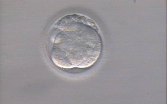
Figure 300
A poor quality morula with several cells and fragments excluded from the main mass of compacted cells.
-
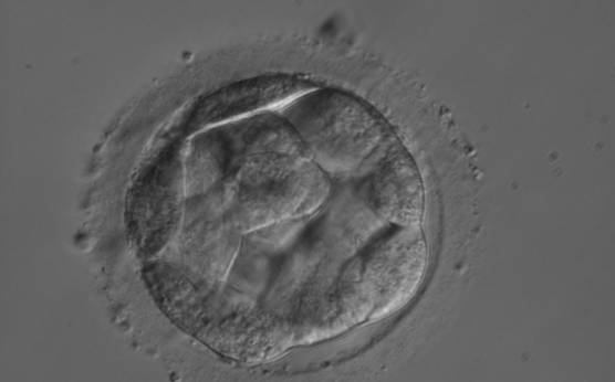
Figure 301
Embryo showing early cavitation with an initial blastocoele cavity beginning to appear.
-
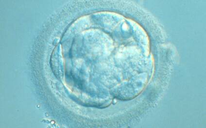
Figure 302
Embryo showing early cavitation. An initial blastocoele cavity is beginning to appear. The ZP is broken after blastomere biopsy.