Chapter 4
b. ICM morphology
-
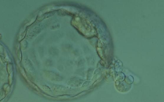
Figure 364
Hatching blastocyst (Grade 5:1:1) with two separate ICMs that appear to be connected to each other at the 2 o'clock position in this view. One ICM appears to be smaller than the other and is beginning to herniate through a breach in the ZP at the 4 o'clock position along with several TE cells. The blastocyst was transferred and failed to implant.
-
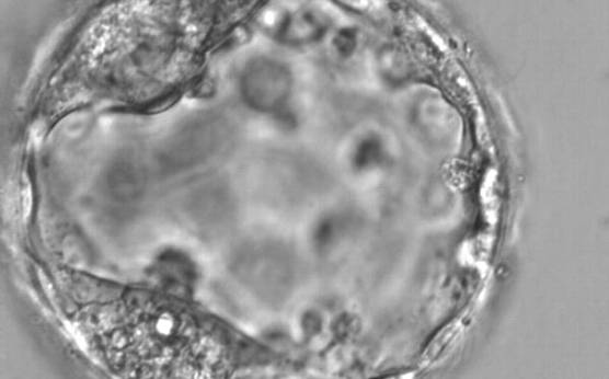
Figure 363
Expanded blastocyst (Grade 4:1:1) with two distinct ICMs at the 7 and 11 o'clock positions in this view. Both ICMs are of reasonable size and compact.
-
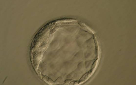
Figure 362
Blastocyst (Grade 3:1:1) with a crescent-shaped ICM at the 10 o'clock position in this view. The blastocyst was transferred but the outcome is unknown.
-
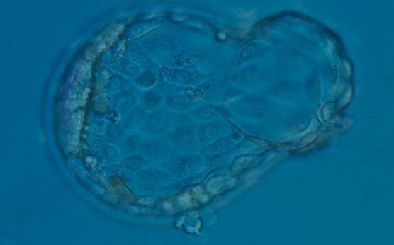
Figure 360
Hatching blastocyst (Grade 5:1:1) with a compact crescent-shaped ICM flattened to the TE at the 9 o'clock position in this view. There is a large amount of TE herniating from a breach in the ZP at the 3 o'clock position and a second smaller site of herniation at the 6 o'clock position. The blastocyst developed following ICSI and so the breach in the ZP at the 6 o'clock position may be a result of the injection site. This blastocyst did not result in a pregnancy following transfer.
-
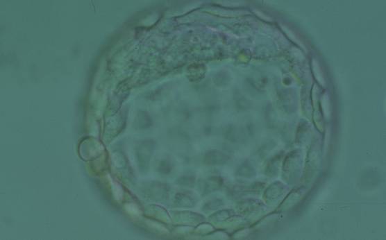
Figure 361
Hatching blastocyst (Grade 5:1:1) showing a large crescent-shaped ICM at the 12 o'clock position in this view. The blastocyst was transferred and implanted.
-
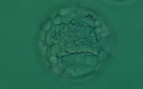
Figure 359
Expanded blastocyst (Grade 4:1:1) with a large stellate ICM present at the base of the blastocyst in this view. The blastocyst was transferred and implanted.
-
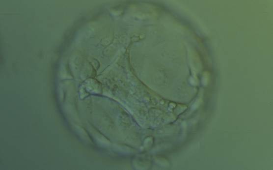
Figure 358
Hatching blastocyst (Grade 5:1:2) with a large stellate ICM present at the base of the blastocyst in this view. The ICM seems to be connected to the TE cells with short triangular cytoplasmic strings or bridges. The blastocyst was transferred but failed to implant.
-
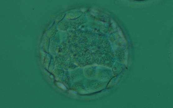
Figure 357
Expanded blastocyst (Grade 4:1:1) with a large stellate ICM flattened at the base of the blastocyst in this view. The blastocyst was transferred and implanted.
-
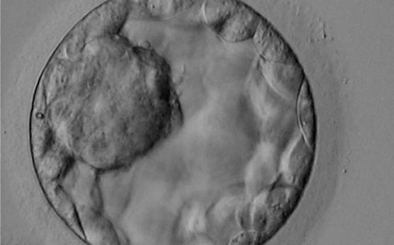
Figure 356
Blastocyst (Grade 3:1:1) showing a very large, mushroom-shaped ICM at the 10 o'clock position in this view and made up of many cells that are tightly compacted. The blastocyst was transferred and resulted in the delivery of a healthy girl.
-
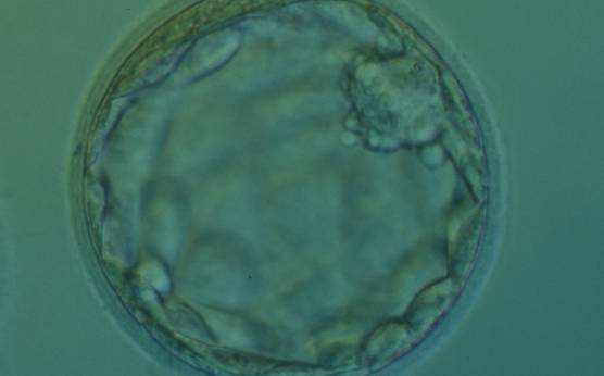
Figure 355
Expanded blastocyst (Grade 4:2:1) with a small, mushroom-shaped compact ICM. There is some cellular debris present in the space between the ZP and the TE at the 10–12 o'clock positions. The blastocyst was transferred but the outcome is unknown.
-
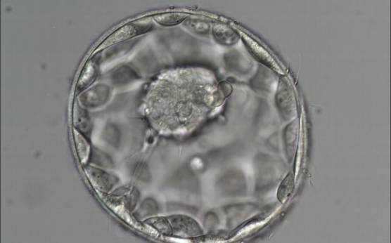
Figure 354
Good quality expanded blastocyst (Grade 4:1:1) with a large mushroom-shaped ICM. There appears to be cytoplasmic strings extending from the ICM to the TE. The blastocyst was transferred and implanted.
-
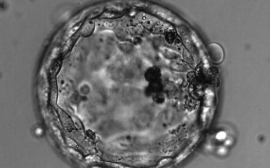
Figure 353
Hatching blastocyst (Grade 4:3:1) with no clearly identifiable ICM but a TE that is made up of many cells forming a cohesive epithelium. Dark, degenerate cells are present toward the 3 o′clock position in this view.
-
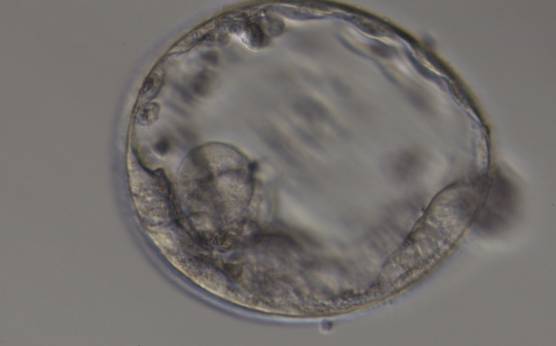
Figure 352
Expanded blastocyst (Grade 4:3:3) with no clearly identifiable ICM. TE is made up of a few, sparse cells that do not form a cohesive epithelium.
-
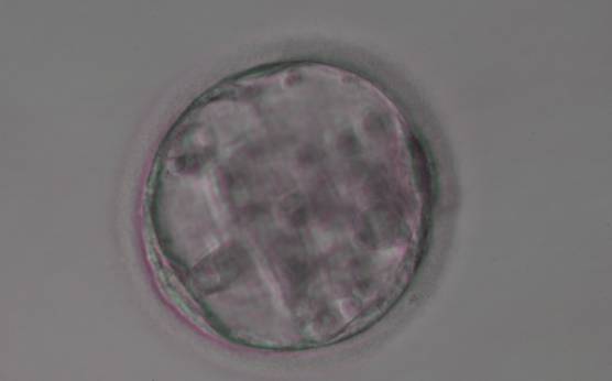
Figure 351
Blastocyst (Grade 3:3:3) with no clearly identifiable ICM and sparse TE that does not form a cohesive epithelium.
-
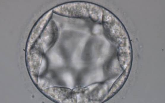
Figure 350
Blastocyst (Grade 3:3:2) with no clearly identifiable ICM and with TE cells that in places are quite large and stretch over great distances to reach the next cell.
-
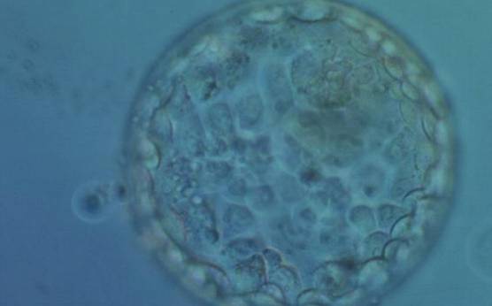
Figure 349
Hatching blastocyst (Grade 5:2: 1) showing a compact ICM toward the 1 o′clock position in this view. The ICM is very small and made up of few cells relative to the size of the blastocyst. There are a few small, dark degenerative foci in the TE cells. The blastocyst was transferred but failed to implant.
-
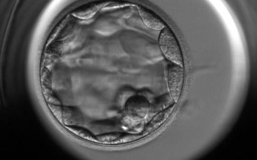
Figure 347
Blastocyst (Grade 3:2:1) showing a compact ICM at the 5 o'clock position in this view. The ICM is very small and made up of only a few cells. The blastocyst was transferred but the outcome is unknown.
-
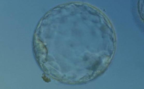
Figure 348
Hatching blastocyst (Grade 5:2:1) showing a compact ICM at the 8 o′clock position in this view. The ICM is small and flattened and made up of few cells relative to the size of the blastocyst. The blastocyst was transferred but the outcome is unknown.
-
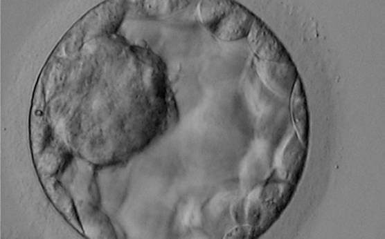
Figure 344
Blastocyst (Grade 3:1:1) showing a very large, mushroom-shaped ICM at the 10 o′clock position in this view. The ICM is made up of many cells that are tightly compacted. The blastocyst was transferred and resulted in the delivery of a healthy girl.
-
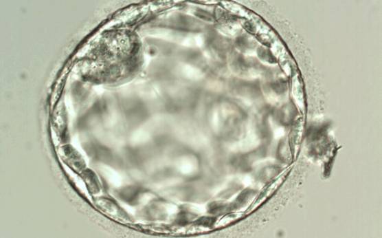
Figure 346
Blastocyst (Grade 3:2:1) showing a compact ICM at the 11 o'clock position in this view. The ICM is small relative to the diameter of the blastocyst and probably made up of few cells. The blastocyst was transferred but failed to implant.
-
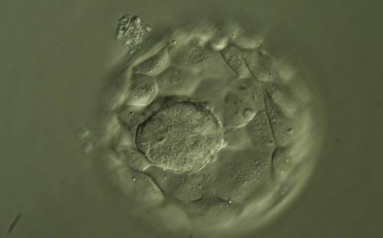
Figure 345
Hatching blastocyst (Grade 5:1:1) showing a large ICM at the base of the blastocyst in this view. The ICM is made up of many cells that are tightly compacted. The blastocyst was transferred but the outcome is unknown.
-
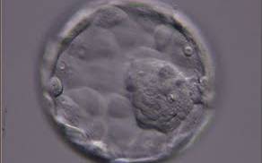
Figure 342
Expanded blastocyst (Grade 4:1:1) showing a large ICM at the 4 o'clock position in this view. The ICM is made up of many cells and is very compact. The blastocyst was transferred but the outcome is unknown.
-
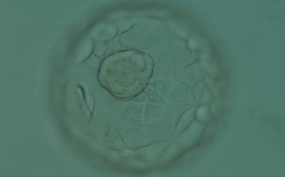
Figure 343
Expanded blastocyst (Grade 4:1:1) showing a large ICM at the base of the blastocyst in this view. The ICM is made up of many cells that are tightly compacted. The blastocyst was transferred but the outcome is unknown.