Chapter 4
d. Cellular degeneration in blastocysts
-
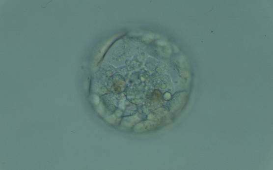
Figure 380
Blastocyst (Grade 3:1:1) with a compact ICM at the base of the blastocyst that is associated with several granular cellular fragments and two darker foci of cell degeneration. The TE cells appear to be healthy and form a cohesive epithelium. The blastocyst was transferred but failed to implant.
-
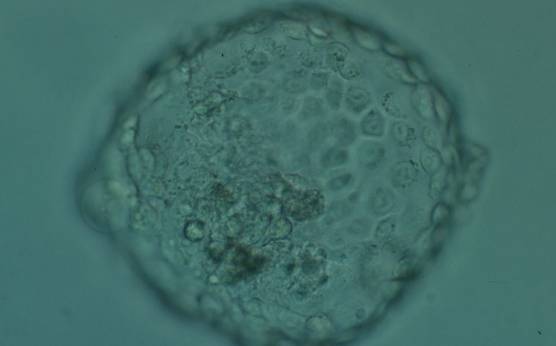
Figure 381
Hatched blastocyst (Grade 6:1:1) showing many cells in the TE making a cohesive epithelium. Many of the TE cells contain dark granules. The ICM is not clearly seen in this view and there are several degenerative foci (dark cells) associated with the ICM and polar TE. The blastocyst was transferred and implanted.
-
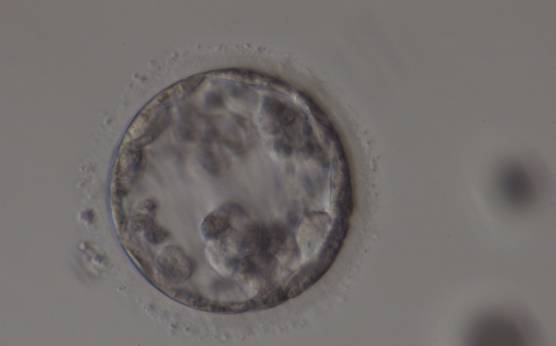
Figure 382
Poor quality, degenerating, early blastocyst in which the majority of cells are showing dark, degenerative changes.
-
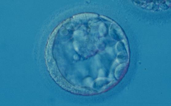
Figure 383
Blastocyst (Grade 3:1:2) with a large, mushroom-shaped ICM toward the 1 o'clock position in this view. From the 6 o'clock to the 3 o'clock position there is a significant amount of cellular debris, both large and small, between the mural TE and the ZP.
-
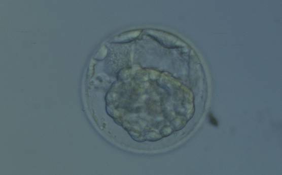
Figure 384
Hatching blastocyst that has collapsed into a dense mass of cells making it impossible to evaluate the ICM and TE cells. Several cells not participating in blastocyst formation can be seen in the PVS.
-
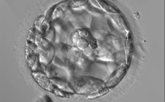
Figure 385
An early blastocyst with a large blastocoel cavity. Note the large cellular debris sequestered into the PVS which does not take part in the blastocyst formation.
-
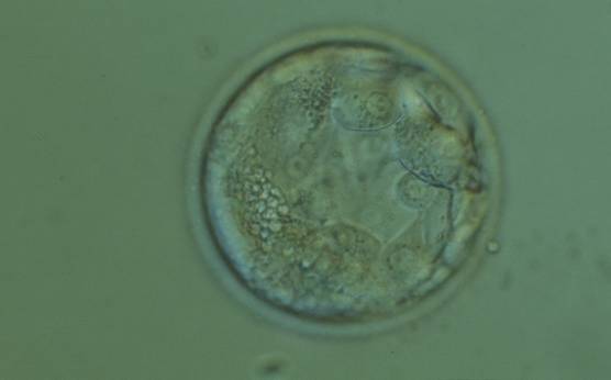
Figure 386
Early blastocyst (Grade 2) in which the ICM is not as yet clearly discernible. Many of the TE cells contain dark granules. In addition there is a large number of small fragments which can be best seen between the 8 and 9 o'clock positions in this view. The blastocyst was transferred but the outcome is unknown.
-
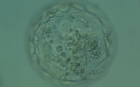
Figure 387
Expanded blastocyst (Grade 4:1:1) in which the ICM is not clear in this view. The TE cells vary in size but form a cohesive epithelium. There are several small- to medium-sized fragments present inside the blastocoel cavity, some of which are quite dark. The blastocyst was transferred but failed to implant.
-
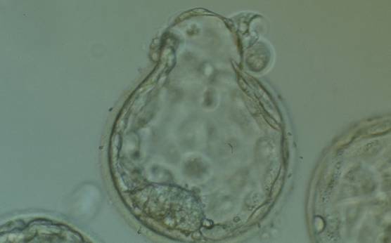
Figure 388
Hatching blastocyst (Grade 5:1:1) in which a large, compact ICM can be seen at the 7 o'clock position and the herniating TE can be seen at the 12 o'clock position in this view. Several large fragments can be observed inside the blastocoel cavity. The blastocyst failed to implant after transfer.