Chapter 4
f. Other morphological features
-
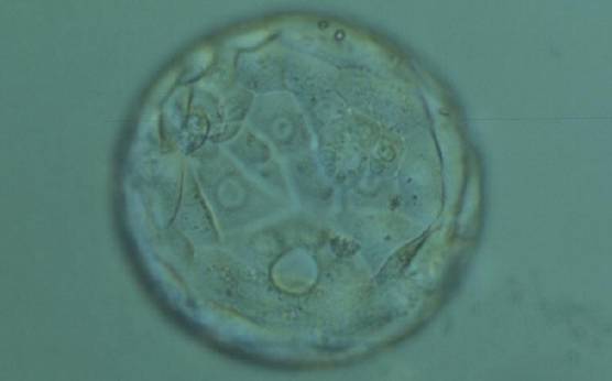
Figure 392
Expanded blastocyst (Grade 4:3:2) in which a large vacuole can be seen at the 6 o'clock position between TE cells. The blastocyst was transferred but the outcome is unknown.
-
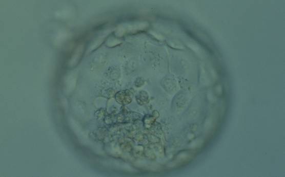
Figure 393
Hatching blastocyst (Grade 5:1:1) showing a compact ICM at the 6 o'clock position which is associated with several cellular fragments. The TE is made up of many cells that form a cohesive epithelium but there are two medium-sized vacuoles within TE cells abutting the ICM toward the 9 o'clock position. The blastocyst was transferred but failed to implant.
-
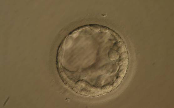
Figure 394
Early blastocyst (Grade 2) with large vacuolization of the TE distinct from the blastocoel cavity at the 10 o'clock position in this view. The blastocyst was transferred but the outcome is unknown.
-
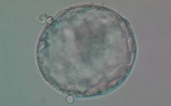
Figure 395
Early hatching blastocyst (Grade 5:1:1) which is beginning to hatch naturally from two different small breaches in the ZP. The ICM is large and compact and the TE cells are many and form a cohesive epithelium. The blastocyst was transferred but the outcome is unknown.
-
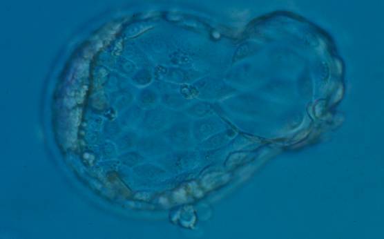
Figure 396
Hatching blastocyst (Grade 5:1:1) with a large amount of TE herniating from a breach in the ZP at the 3 o'clock position and a second smaller site of herniation at the 6 o'clock position. The blastocyst developed following ICSI and so the breach in the ZP at the 6 o'clock position may be a result of the injection site. This blastocyst did not result in a pregnancy following transfer.
-
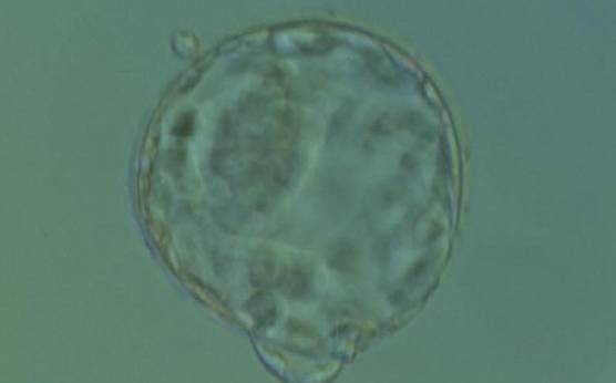
Figure 397
Hatching blastocyst (Grade 5:1:1) with two distinct points of natural hatching, one at the 11 o'clock and one at the 6 o'clock position. The ICM is large and compact and the TE cells are many and form a cohesive epithelium. There is a significant difference in the diameter of the two breaches in the ZP. The blastocyst developed following standard insemination and so the smaller diameter breach is not a result of sperm injection. The blastocyst was transferred but failed to implant.