The Oocyte
a. Cumulus-Enclosed Oocytes
a. Cumulus-Enclosed Oocytes
-
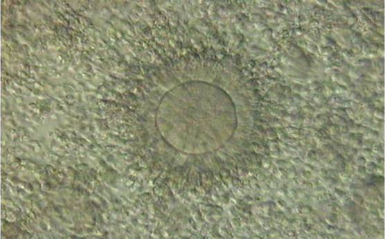
Figure 1. Cumulus–oocyte complex obtained following ovarian stimulation. The oocyte is typically surrounded by an expanded cumulus corona cell complex. Note the outer CCs separated from each other by extracellular matrix and the corona cells immediately adjacent to the oocyte becoming less compact and radiating away from the ZP. PB1 is located at the 1 o'clock position.
-
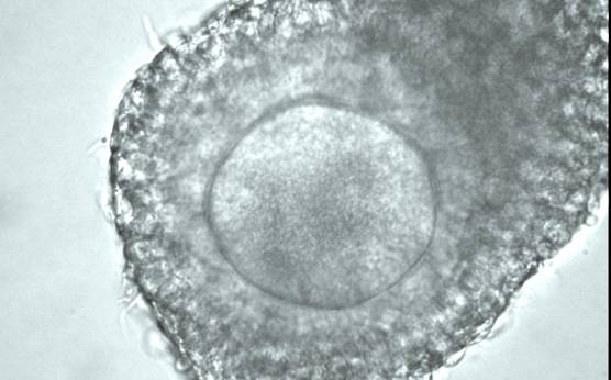
Figure 2
A cumulus–oocyte complex recovered from an IVM cycle showing an oocyte surrounded by unexpanded, compact cumulus and corona cells.
-
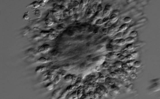
Figure 3. A cumulus–oocyte complex recovered from an IVM cycle. The immature GV oocyte is surrounded by compact GCs. The nucleolus in the GV is visible at the 10 o'clock position.
-
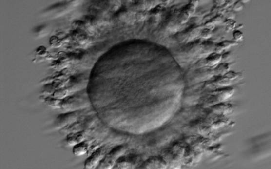
Figure 4. A cumulus–oocyte complex recovered from an IVM cycle. The immature oocyte has a GV at the 3 o'clock position with the nucleolus towards the centre of the oocyte. Compact GCs surround the oocyte.
-
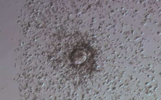
Figure 7
Denudation sequences of a mature oocyte.
a) Cumulus–corona oocyte complex before the denudation process with compact, non-radiating CCs. (100× magnification).
-
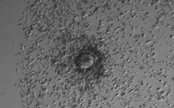
Figure 5. A cumulus oocyte complex at low magnification. The oocyte is surrounded by an expanded cumulus–corona cell complex clearly showing the separation of individual CCs due to the accumulation of hyaluronic acid in the extracellular space.
-
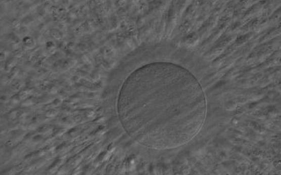
Figure 6. A cumulus–oocyte complex obtained following ovarian stimulation. An expanded cumulus corona cell complex surrounds the oocyte with the outer CCs separated from each other by extracellular matrix. The CCs immediately adjacent to the oocyte become less compact and radiate away from the ZP. The oocyte in the figure can be clearly seen through the surrounding cells at high magnification and no polar body can be seen in the PVS despite the mature status of the cumulus–corona cells.
-
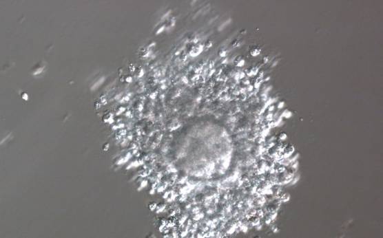
Figure 7
Denudation sequences of a mature oocyte.
b) Oocyte surrounded by corona cells during hyaluronidase treatment (200× magnification).
-
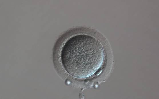
Figure 7
Denudation sequences of a mature oocyte.
c) Denuded oocyte after mechanical stripping, a visible polar body is present in the PVS (200× magnification).
-
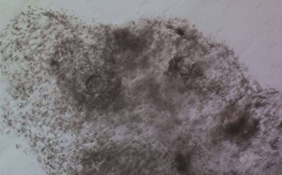
Figure 8
Denudation sequences of a mature oocyte
a) Cumulus–corona oocyte complex before the denudation process (100× magnification). CCs are abundant.
-
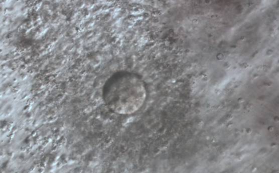
Figure 8
Denudation sequences of a mature oocyte.b) Oocyte surrounded by corona cells during hyaluronidase treatment (200× magnification). Many CCs are still present, but the mature oocyte is already visible with PB1 at the 7 o'clock position.
-
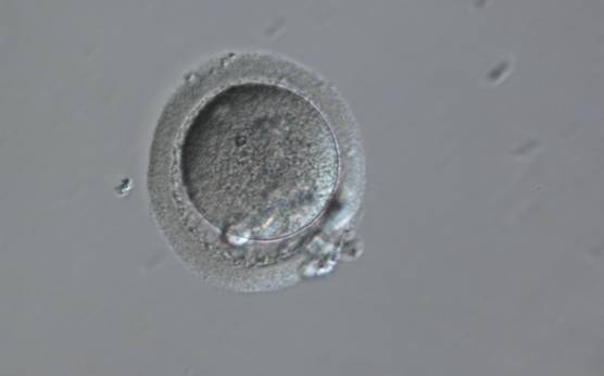
Figure 8
Denudation sequences of a mature oocyte.
c) Denuded oocyte after mechanical stripping, a visible polar body is present in the PVS (200× magnification). -
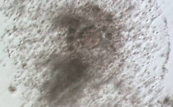
Figure 9
Denudation sequences of a mature oocyte.a) Cumulus–corona oocyte complex before the denudation process with compact, non-radiating CCs. (100× magnification).
-
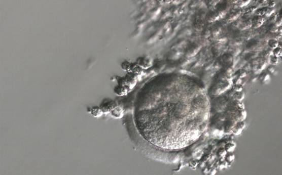
Figure 9
Denudation sequences of a mature oocyte.b) Oocyte surrounded by corona cells during hyaluronidase treatment (200× magnification).
-
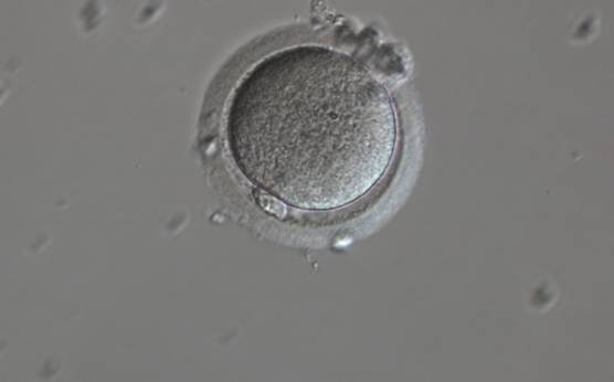
Figure 9
Denudation sequences of a mature oocyte.c) Denuded oocyte after mechanical stripping, a visible polar body is present in the PVS (200× magnification).