Chapter 1
b. Oocyte maturation stage
-
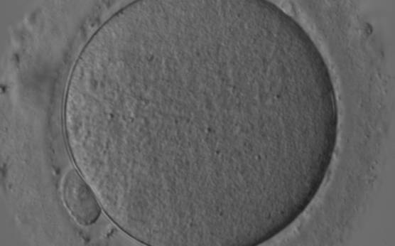
Figure 10
Denuded MII oocyte; an intact PBI is clearly visible in the PVS (400× magnification).
-
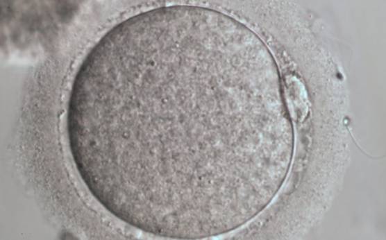
Figure 11
Denuded MII oocyte; the PBI is clearly visible in the narrow PVS (400× magnification).
-
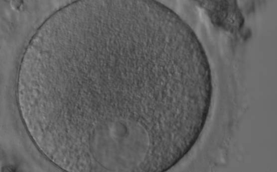
Figure 12
Denuded GV oocyte. A typical GV oocyte with an eccentrically placed nucleus and a prominent single nucleolus (400× magnification).
-
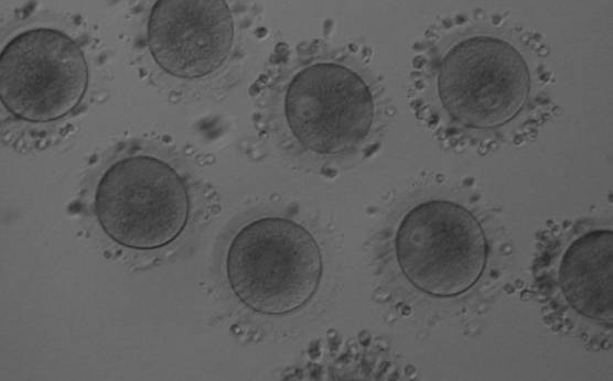
Figure 13
Denuded GV oocytes. Several GV with the organelles condensed centrally within the cytoplasm (200× magnification).
-
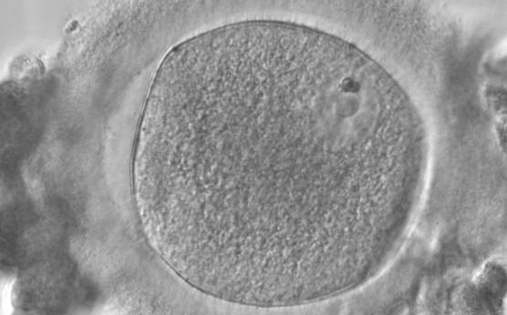
Figure 14
Denuded GV oocyte. A GV oocyte that is possibly approaching GVBD as the nuclear membrane is not distinct over its entirety (400× magnification).
-
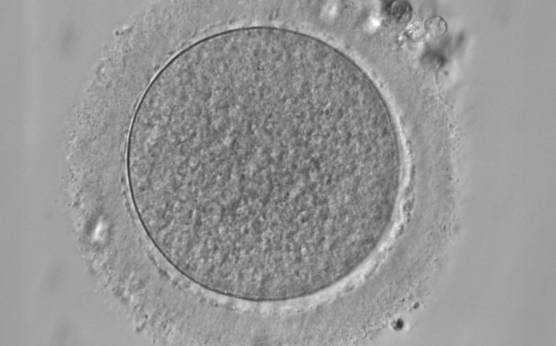
Figure 15
Denuded MI oocyte. This oocyte has no visible nucleus and has not as yet extruded the PBI (400× magnification). PVS is typically narrow.
-
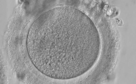
Figure 16
Denuded MI oocyte with no visible nucleus and no PBI (400× magnification). Some CCs are still tightly adhered to the ZP.
-
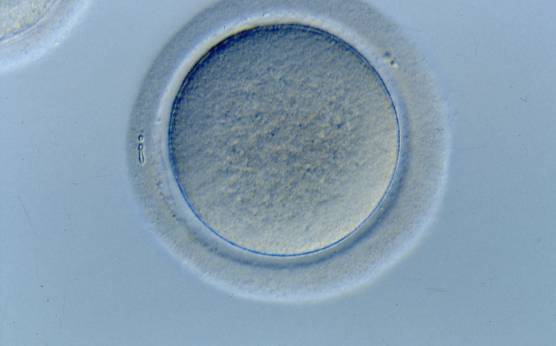
Figure 17
Denuded MI oocyte without a visible nucleus or an extruded PBI (400× magnification).
-
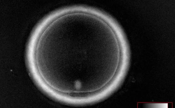
Figure 18
MII oocyte visualized using polarized light microscopy (400× magnification). The polar body is present at the 6 o'clock position in the PVS, and the MS of the second meiotic division is visible in the cytoplasm perfectly aligned to PB1 position. This is a fully mature MII oocyte.
-
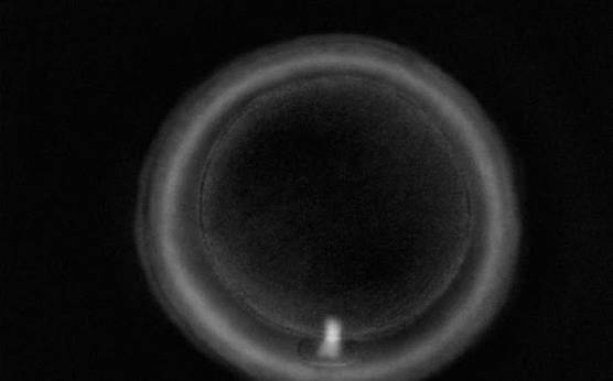
Figure 19
Telophase I oocyte visualized using polarized light microscopy (400× magnification). PB1 is present in the PVS; however, the MS can be seen between PB1 and the oocyte cytoplasm indicating that this oocyte is still completing the first meiotic division. This is not yet a fully mature MII oocyte.
-
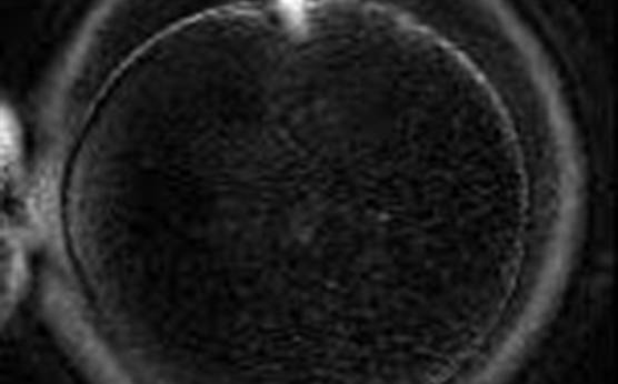
Figure 20
Telophase I oocyte visualized using polarized light microscopy (400× magnification). The MS can be seen between PB1 and the oocyte cytoplasm indicating that the first meiotic division is not yet completed.
-
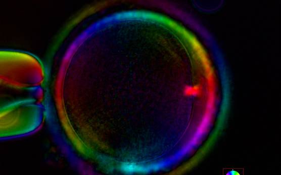
Figure 21
Telophase I oocyte visualized using polarized light microscopy (400× magnification). PB1 is present in the PVS at the 3 o'clock position; however, the MS is still bridging PB1 and the oocyte cytoplasm indicating that this oocyte is not yet a fully mature MII oocyte.
-
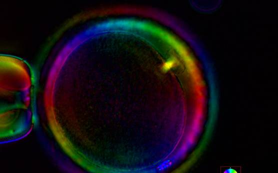
Figure 22
Telophase I oocyte visualized using polarized light microscopy (400× magnification). The MS can be seen between PB1 and the oocyte cytoplasm indicating that this oocyte is still completing the first meiotic division despite the extrusion of PB1 in the PVS.
-
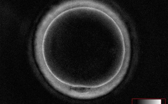
Figure 23
Interphase oocyte (between the first and second meiotic division; Prophase II) visualized using polarized light microscopy (400× magnification). PB1 is present in the PVS at the 6 o'clock position; however, the MS of the second meiotic division is not yet visible in the cytoplasm. This is not yet a fully mature MII oocyte.