The Oocyte
C. Oocyte size and shape
C. Oocyte size and shape
-
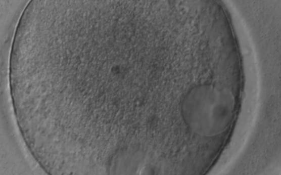
Figure 25
Giant oocyte with two apparent GVs (in an eccentric position). This is a tetraploid oocyte originating from the fusion of two separate oocytes (400× magnification).
-
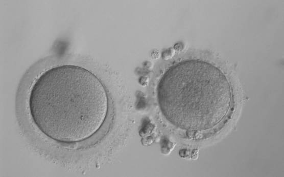
Figure 24
Small MII oocyte (right) next to a normal-sized MII oocyte (left) from the same cohort (200× magnification).
-
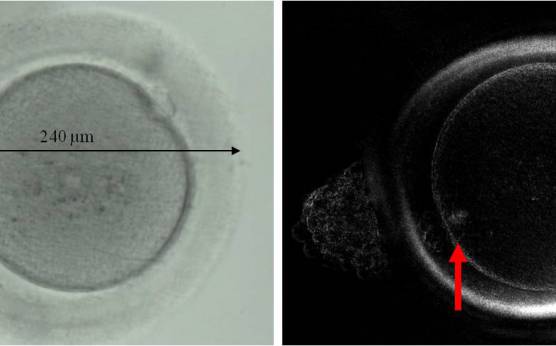
Figure 27
Giant MII oocyte visualized using bright field (left) and polarized light microscopy (right). The oocyte contains two distinct polar bodies and two distinct MSs at opposing poles of the oocyte.
-
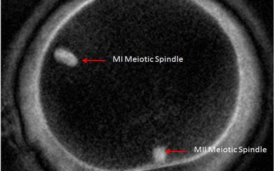
Figure 28
Giant MII oocyte visualized at high power using polarized light microscopy. The two distinct MSs can be observed in the cytoplasm. One MS is from the MI (10 o'clock position) and the other is from MII (6 o'clock position; 400× magnification).
-
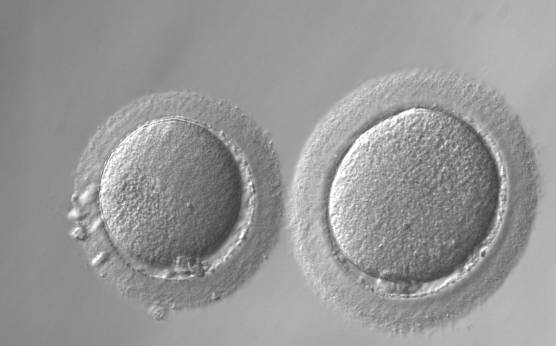
Figure 29
Giant oocyte (right) next to normal-sized oocyte (left; 200× magnification).
-
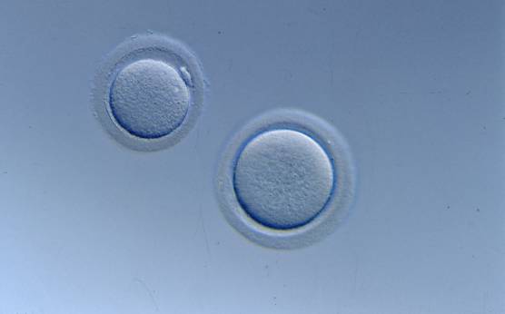
Figure 30
Giant oocyte (right) next to normal-sized oocyte (left; 200× magnification). No PB1 is visible in the giant oocyte.
-
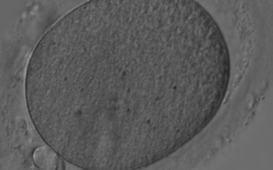
Figure 31
Elongated MII oocyte inside an elongated ZP. Note the PVS appears relatively normal (400× magnification).
-
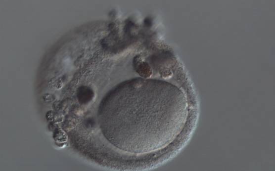
Figure 32
Elongated MII oocyte within a grossly distended and irregular ZP (200× magnification).
-
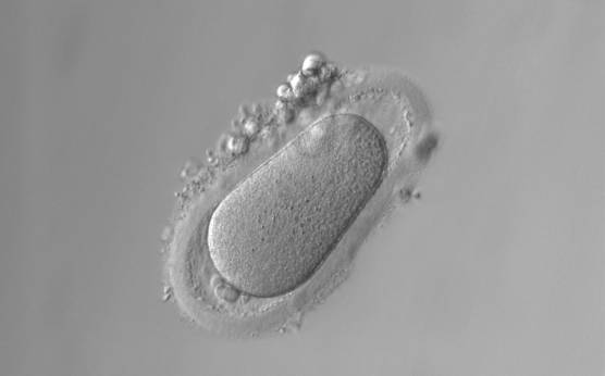
Figure 33
Ovoid MII oocyte. Note the ZP is also ovoid in appearance and the PVS is enlarged at both poles (200× magnification).
-
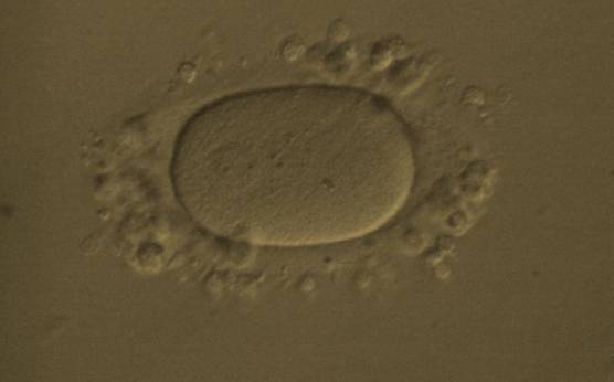
Figure 34
Ovoid MII oocyte. Note the ZP is also ovoid in appearance permitting the PVS to remain relatively normal (200× magnification).
-
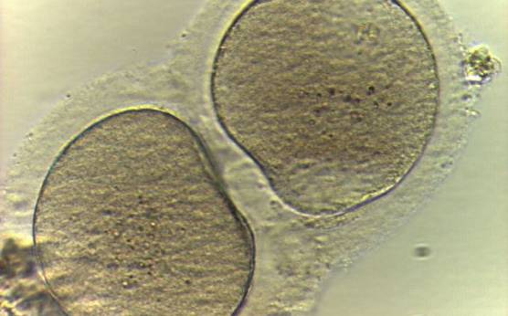
Figure 35
Two oocytes enclosed within a single ZP (400× magnification).
-
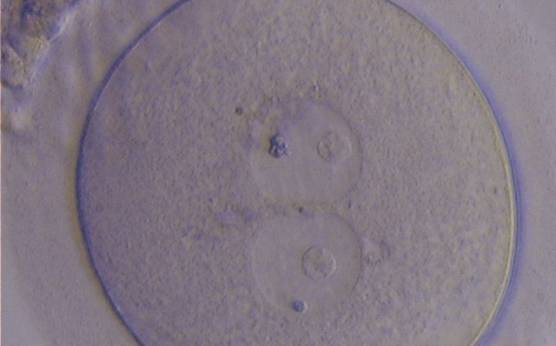
Figure 26
Giant oocyte with two apparent GVs (centrally located and juxtapposed). This tetraploid oocyte originates from the fusion of two separate oocytes and is usually tetraploid (400× magnification).