The Oocyte
Cytoplasmic Features
Cytoplasmic Features
-

Figure 36
Normal homogenous cytoplasm in an MII oocyte (400× magnification).
-
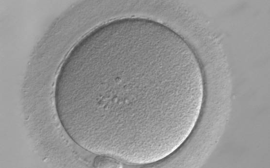
Figure 37
Normal homogenous cytoplasm in an MII oocyte (400× magnification). ZP is dense and homogeneous.
-
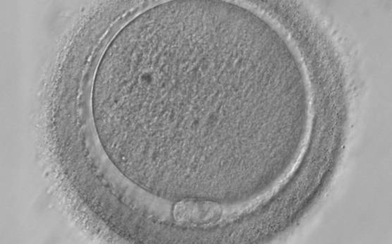
Figure 38
MII oocyte with granular cytoplasm (400× magnification). ZP is thicker in the lower-left part of the oocyte in this view.
-
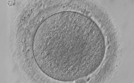
Figure 39
MII oocyte with granular cytoplasm (400× magnification). ZP is also abnormal with marked differences in thickness.
-
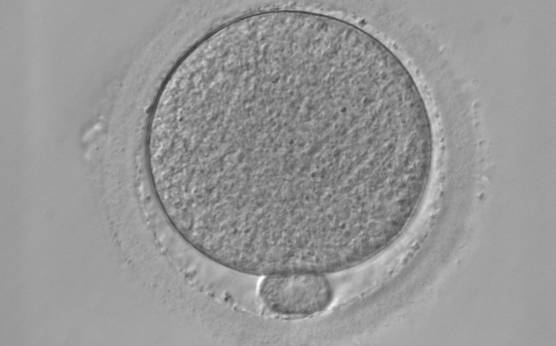
Figure 40
MII oocyte with granular cytoplasm. This figure can be considered as a slight deviation from normal homogeneous cytoplasm (400× magnification).
-
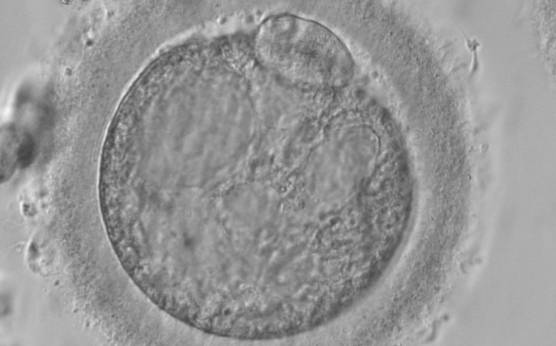
Figure 41
MII oocyte with a high degree of cytoplasmic granularity/degeneration (400× magnification). PB1 is larger than normal size.
-
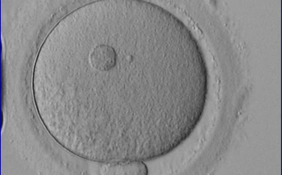
Figure 42
A large refractile body can be seen within the oocyte cytoplasm (400× magnification).
-
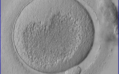
Figure 43
MII oocyte showing a very large centrally located granular area occupying the majority of the cytoplasm. This granularity is typical of organelle clustering.
-
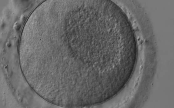
Figure 44
MII oocyte showing a large centrally located granular area in the cytoplasm denoting organelle clustering.
-
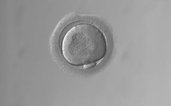
Figure 45
MII oocyte showing organelle clustering forming a large centrally located granular area in the cytoplasm. ZP has an irregular shape.
-
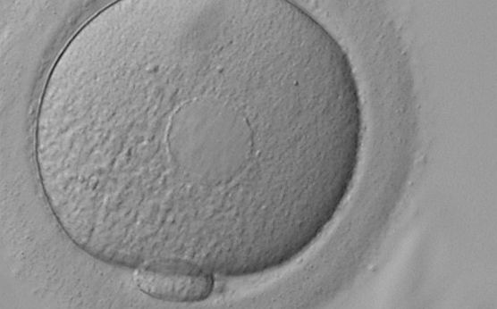
Figure 46
MII oocyte showing plaques of dilated SER discs in the cytoplasm (400× magnification). The SER discs can be clearly distinguished from the fluid-filled vacuoles pictured in Figs 48–51. The cytoplasm looks heterogeneous with granularity (on the left) and areas devoid of organelles toward the 12 o'clock position in this view.
-
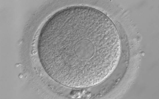
Figure 47
MII oocyte showing plaques of dilated SER discs in the cytoplasm (400× magnification). The PVS is enlarged and PB1 is fragmented.
-
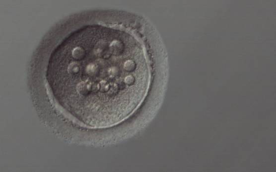
Figure 48
Vacuolated oocyte. Depicts an oocyte with several small vacuoles distributed throughout the oocyte cytoplasm (200× magnification).
-
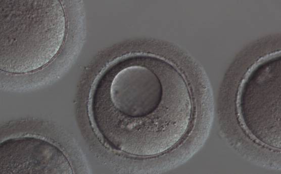
Figure 49
Vacuolated oocyte. This oocyte has a single large vacuole occupying almost half of the oocyte cytoplasm (200× magnification).
-
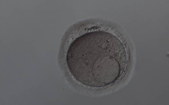
Figure 50
Vacuolated oocyte. This oocyte has a single large vacuole in a dark, granular cytoplasm (200× magnification).
-
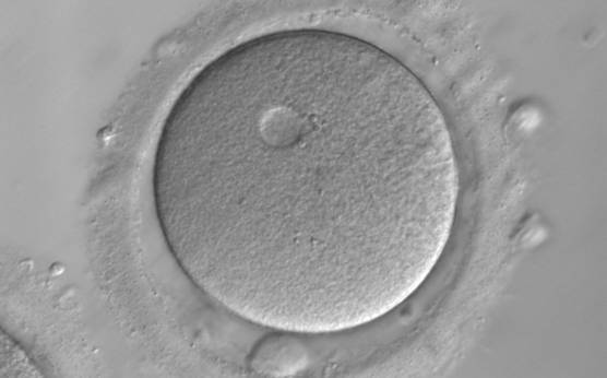
Figure 51
Vacuolated oocyte. Depicts an oocyte with a single small vacuole in the oocyte cytoplasm (400× magnification).
-
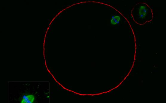
Figure 52
MII oocyte with a normal-shaped MS observed using confocal microscopy.
-
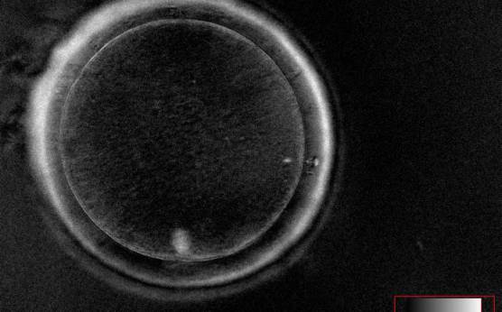
Figure 53
MII oocyte observed using polarized light microscopy with a visible MS just below PB1 (400× magnification).
-
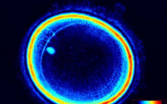
Figure 54
MII oocyte observed using polarized light microscopy with a visible MS near to PB1 (400× magnification).
-
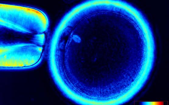
Figure 55
MII oocyte observed using polarized light microscopy with a visible MS just below PB1 that appears fragmented (two fragments). (400× magnification).
-
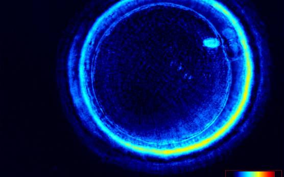
Figure 56
MII oocyte observed using polarized light microscopy. The MS is clearly visible near to PB1 (400× magnification).
-
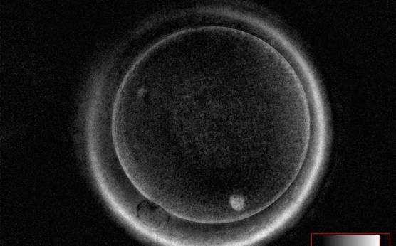
Figure 57
MII oocyte observed using polarized light microscopy with a visible MS slightly dislocated from PB1 at the 7 o'clock position in this view (400× magnification).
-
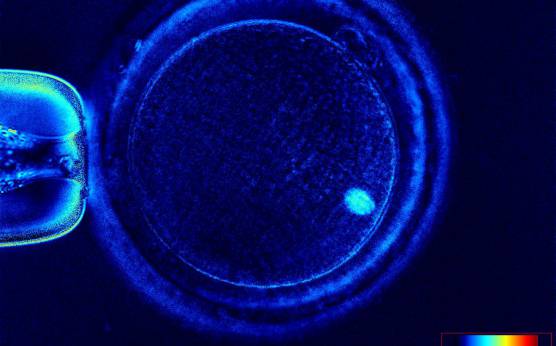
Figure 58
MII oocyte observed using polarized light microscopy with a visible MS dislocated about 80° from PB1 at the 1 o'clock position in this view (400× magnification).
-
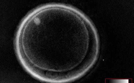
Figure 59
MII oocyte observed using polarized light microscopy with a visible MS highly dislocated (about 135°) from PB1 at the 6 o'clock position in this view (400× magnification).
-
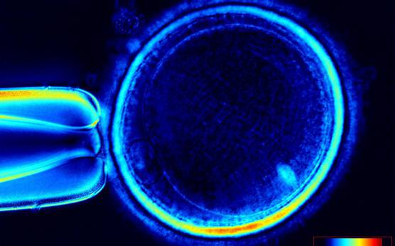
Figure 60
MII oocyte observed using polarized light microscopy with a visible MS highly dislocated (slightly more than 90°) from PB1 at the 8 o'clock position in this view (400× magnification).