The Zygote
Fertilization Assessment
Fertilization Assessment
-
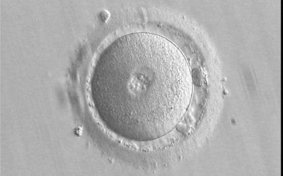
Figure 85
A zygote 16.5 h post-ICSI, having small-sized PNs with scattered NPBs and two visible polar bodies (400× magnification). The zona pellucida (ZP) appears regular; some debris is present in the perivitelline space (PVS). The cytoplasm is homogeneous and displays a clear cortical zone. It was transferred resulting in a clinical pregnancy followed by miscarriage.
-
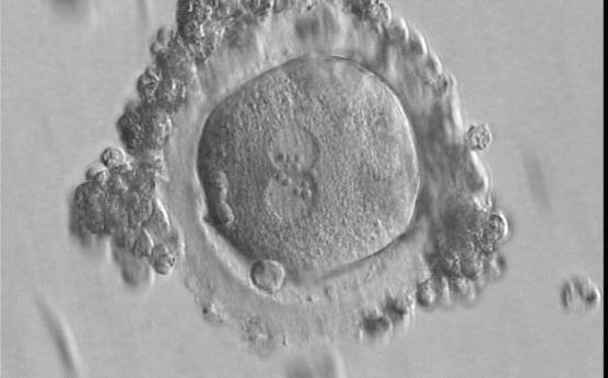
Figure 86
A slightly deformed zygote at 16.5 h after IVF with equal numbers of large-sized NPBs aligned at the PN junction (400× magnification). A great angle separates the two polar bodies. Some granulosa cells surround the ZP. It was transferred but failed to implant.
-
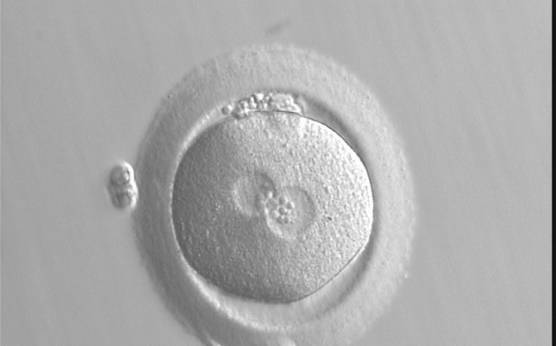
Figure 87
A zygote at 18.5 h generated by standard insemination using frozen/thawed ejaculated sperm (400× magnification). The two PNs are centrally located: one is slightly larger than the other. NPBs are of the same size, but different in number and are aggregated at adjacent borders of each PN. The ZP appears thick. It was transferred but failed to implant.
-
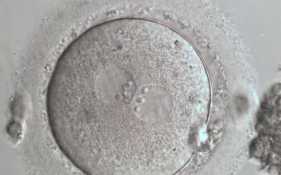
Figure 88
A zygote generated by ICSI with NPBs perfectly aligned at the junction of centrally located and juxtaposed PNs (600× magnification). Fragmented polar bodies are located in the longitudinal axis of the PNs. Category 1 (equivalent to Z1 score; Scott, 2003) was assigned following assessment. Debris appears to be present in the PVS. The cytoplasm is light-coloured with a clear cortical zone. It was transferred and implanted.
-
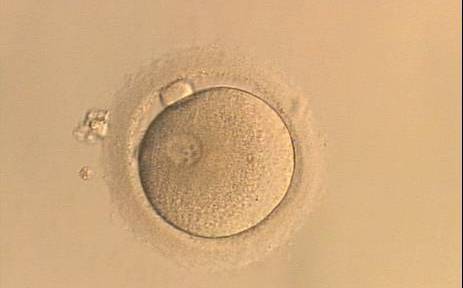
Figure 89
A single pronucleate oocyte displaying only one PN and a single polar body observed 16 h post-ICSI (200× magnification). The observation was repeated 17.5, 20 and 22 h post-ICSI and did not show significant variation in the PN size or position.
-
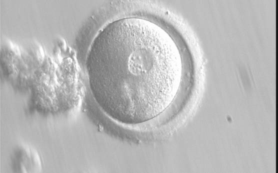
Figure 90
A zygote observed 15 h post-IVF displaying a single, large-sized PN and two polar bodies (400× magnification).
-
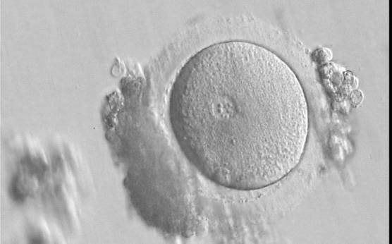
Figure 91
A zygote 17 h post-IVF showing a single PN with NPBs of different size and two polar bodies (400× magnification).
-
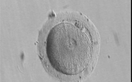
Figure 92
A single, large PN and two polar bodies (partially overlapping) are present in this oocyte observed 16 h and 45 min post-IVF (400× magnification). Four large-sized NPBs are visible. The resulting embryo was transferred, but failed to implant.
-
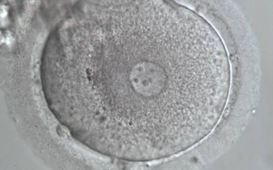
Figure 93
A zygote generated by ICSI showing a single PN and two polar bodies separated by some distance (600× magnification).
-
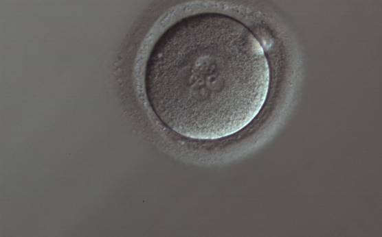
Figure 94
A zygote generated by ICSI displaying four PNs of approximately the same size and two of smaller size (150× magnification). Only one polar body is visible.
-
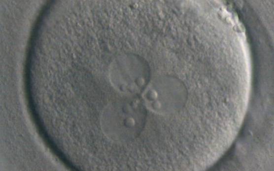
Figure 95
A zygote displaying 3PNs with large-sized NPBs (400× magnification). One of the three PNs is slightly bigger than the others. The zygote was generated by ICSI performed on a giant oocyte.
-
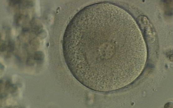
Figure 96
A zygote displaying 3PNs of approximately the same size with large-sized NPBs, partly overlapped and aligned in the middle of the oocyte (400× magnification). It was generated by IVF and shows two polar bodies.
-
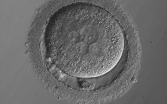
Figure 97
A zygote with 5PNs, halo cytoplasm, fragmented polar bodies, oval shape and dark ZP (400× magnification). It was warmed after vitrification.
-
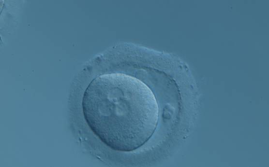
Figure 98
A zygote displaying 3PNs after IVF with a small fragment adjacent to the PNs (200× magnification). There are two polar bodies in a large PVS and a thick ZP.