The Zygote
Pronuclear size
Pronuclear size
-
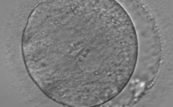
Figure 99
A zygote observed 18 h post-ICSI (400× magnification). The 2PNs are centrally located and juxtaposed in the cytoplasm (peripherally granular), of approximately the same size, and exhibit inequality in the number and size of NPBs. The PN on the right demonstrates fewer but larger nucleoli. Some debris appears to be present in the slightly increased PVS. Transferred on Day 3 (eight cells) along with two other embryos to a patient who delivered a healthy baby boy.
-
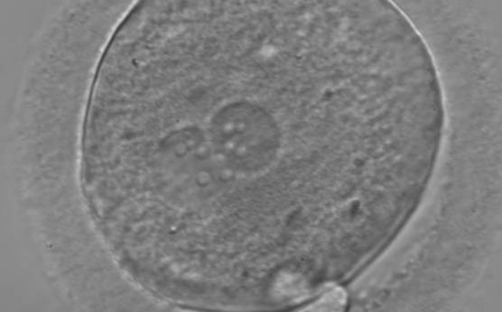
Figure 100
A zygote with equal numbers of large-sized NPBs scattered with respect to the PN junction (400× magnification). PNs are juxtaposed and slightly eccentric. The two polar bodies are located in a plane that is parallel to the longitudinal axis of the PNs.
-
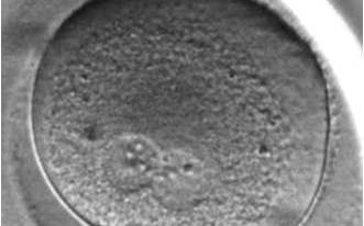
Figure 101(a)
A zygote with changes in PN pattern, particularly with respect to the position in the cytoplasm and the NPBs' location over time at (a) 11.7h after ICSI (400× magnification).
-
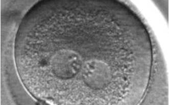
Figure 101(b)
15.4 h after ICSI (400× magnification).
-
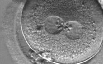
Figure 101(c)
18.3 h after ICSI (400× magnification).
-
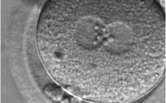
Figure 101(d)
28.3h after ICSI (400× magnification).
-
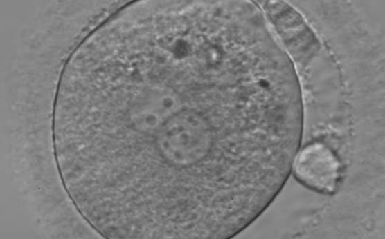
Figure 102
A zygote generated by ICSI and observed 18 h post-insemination (400× magnification). PNs are slightly smaller than normal and exhibit inequality in the number and the distribution of NPBs. Polar bodies are larger than the normal size.
-
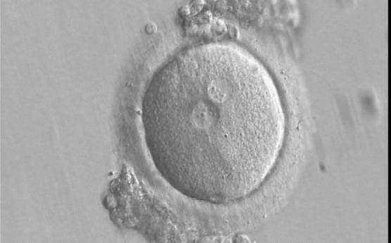
Figure 103
A zygote generated by IVF using frozen/thawed ejaculated sperm and observed 16 h post-insemination (400× magnification). PNs are smaller than normal and are not exactly positioned in the centre of the oocyte. Small-sized NPBs are aligned at the PN junction. It was transferred and implanted.
-
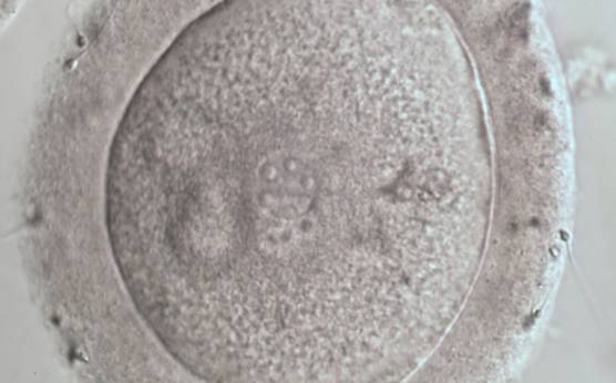
Figure 104
A zygote displaying two small PNs partly overlapping in this view (600× magnification). NPBs are large in size, equal in numbers and scattered in the two PNs. The ZP appears thickened and the PVS almost absent.
-
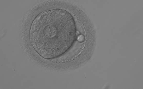
Figure 105
A zygote observed 18 h post-ICSI displaying very unequal-sized juxtaposed PNs, with the smaller PN being less visible (200× magnification). Two polar bodies and small-sized NPBs are scattered in both PNs.
-
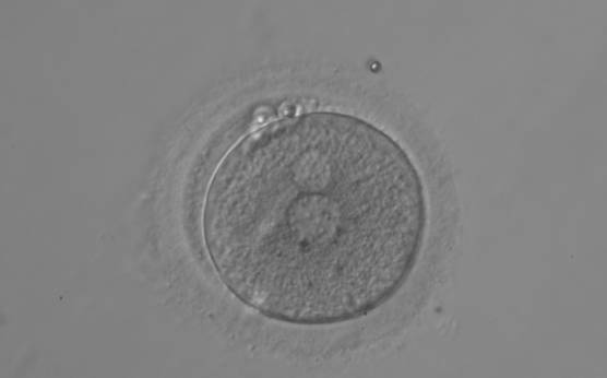
Figure 106
A zygote displaying two polar bodies and unequal-sized juxtaposed PNs and inequality in number and alignment of small-sized NPBs (200× magnification).
-
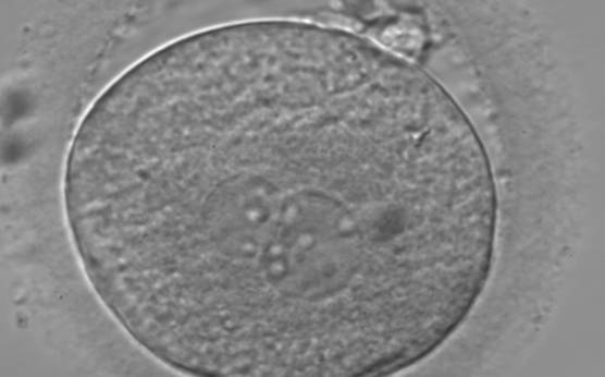
Figure 107
A zygote observed 18 h post-insemination displaying very unequal-sized PNs and inequality in number and alignment of NPBs (400× magnification). It was transferred on Day 3 (seven cells) along with two other embryos. The implantation result is therefore unknown. However, the patient delivered a healthy baby boy.
-
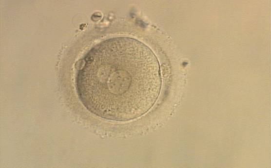
Figure 108
A zygote generated by ICSI with one large and one normal-sized PN (200× magnification). The two polar bodies are at opposite sides of the oocyte.
-
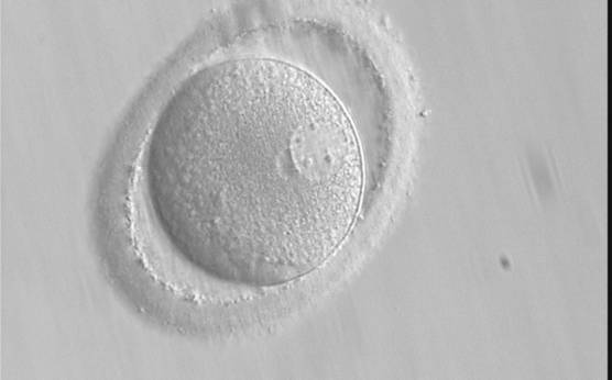
Figure 109
A zygote after PB biopsy showing peripheral PNs that are very different in size with one larger and one smaller than the normal size (400× magnification). The ZP is oval in shape.
-
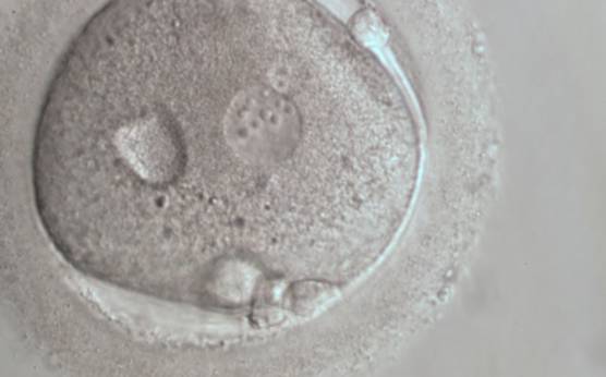
Figure 110
A zygote 18 h post-ICSI displaying very unequal-sized, juxtaposed PNs and inequality in number and alignment of NPBs (600× magnification). A vacuole-like structure is present in the cytoplasm. Fragments are visible in the PVS, not easily discernible from the polar bodies in this view.