The Zygote
Nucleolar precursor bodies
Nucleolar precursor bodies
-
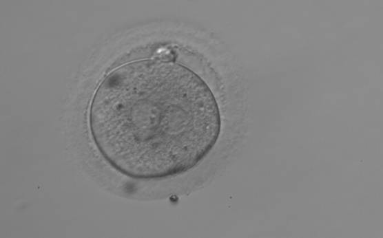
Figure 153
A zygote observed 18 h post-ICSI with NPBs aligned at the PN junction (200× magnification). NPBs differ in number and size between PNs. The polar bodies are overlapped in this view. It was discarded due to subsequent abnormal cleavage.
-
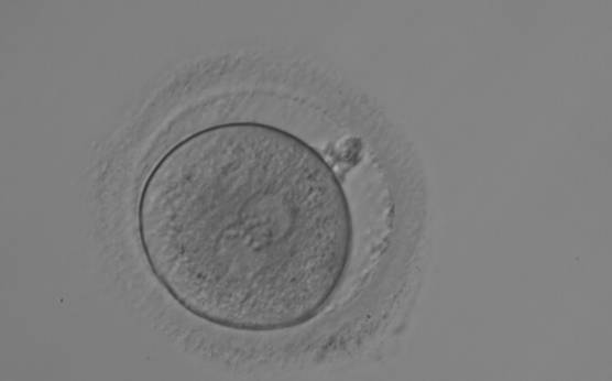
Figure 152
A zygote observed 18 h post-ICSI with equal numbers of NPBs aligned at the PN junction (200× magnification). It shows a large PVS and an irregular ZP. The polar bodies are fragmented and overlapping in this view. It was transferred but the outcome is unknown.
-
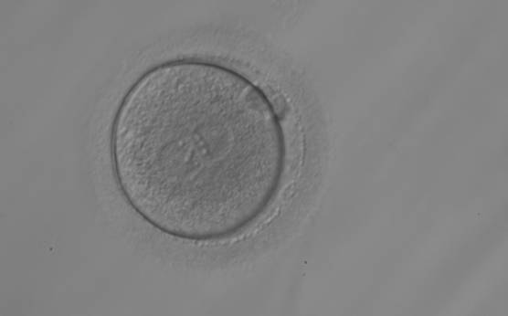
Figure 151
A zygote observed 18 h post-ICSI with equal numbers of NPBs aligned at the PN junction (200× magnification). It was transferred but the outcome is unknown.
-
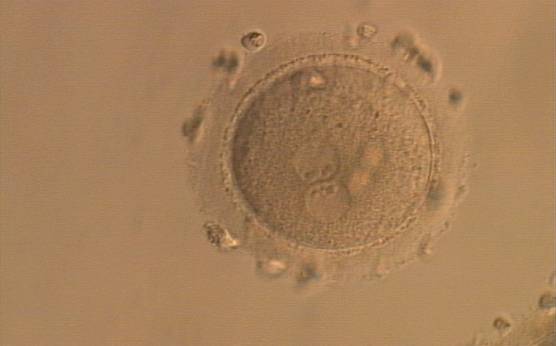
Figure 154
A zygote generated by ICSI, displaying two perfectly juxtaposed PNs within which the NPBs are aligned, have the same size and are similar in number (400× magnification). It was transferred but the outcome is unknown.
-
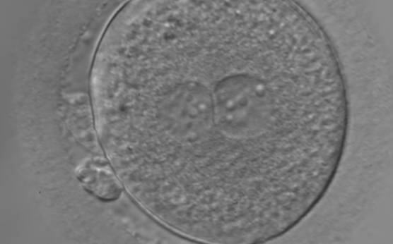
Figure 155
An ICSI zygote with large-sized NPBs scattered with respect to the PN junction (400× magnification). The cytoplasm appears granular. It was transferred but failed to implant.
-
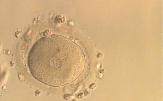
Figure 156
An ICSI zygote with NPBs scattered with respect to the PN junction (400× magnification). Polar bodies are fragmented; there is debris in the PVS. It was transferred but implantation outcome is unknown.
-
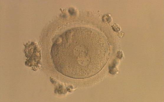
Figure 157
An ICSI zygote displaying partially overlapping PNs in this view. NPBs are scattered in both PNs (400× magnification). It was transferred but the outcome is unknown.
-
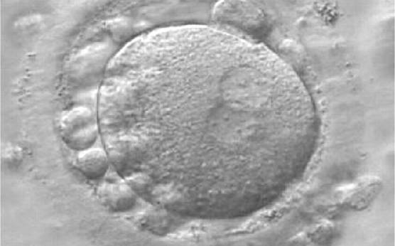
Figure 158
A zygote generated by IVF at 16.40 h post-insemination (400× magnification). It shows small-sized NPBs scattered with respect to the PN junction and a lot of large cellular debris in the PVS. It was discarded.
-
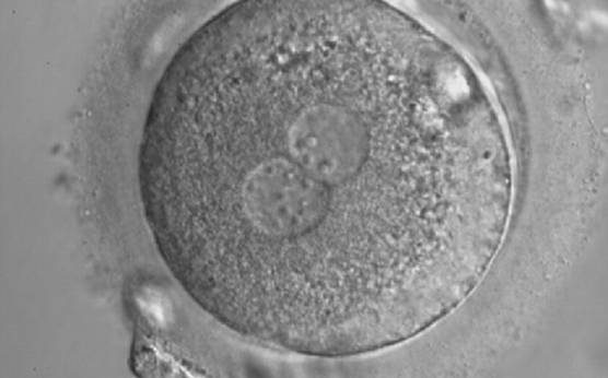
Figure 159
A zygote with inequality in numbers and alignment of NPBs. It was cryopreserved.
-
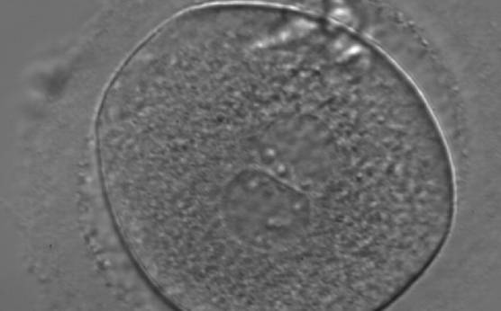
Figure 160
A zygote generated by ICSI with inequality in numbers and alignment of NPBs (400× magnification). NPBs are aligned in one PN and scattered in the other. Due to poor development, it was discarded.
-
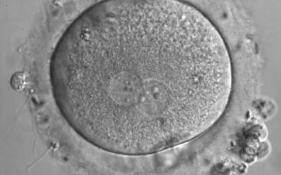
Figure 161
A zygote generated by IVF with inequality in numbers and alignment of NPBs (600× magnification). It was discarded.
-
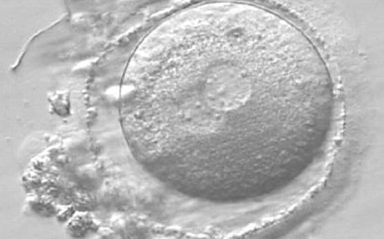
Figure 162
A zygote observed 16 h post-ICSI with small-sized NPBs scattered with respect to the PN junction in both PNs (400× magnification). It has a very thin ZP. It was transferred but failed to implant.
-
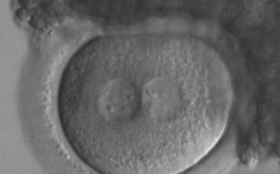
Figure 163
A zygote generated by ICSI with inequality in numbers and alignment of NPBs (200× magnification). Many granulosa cells are adherent to the ZP. It was transferred and implanted.
-
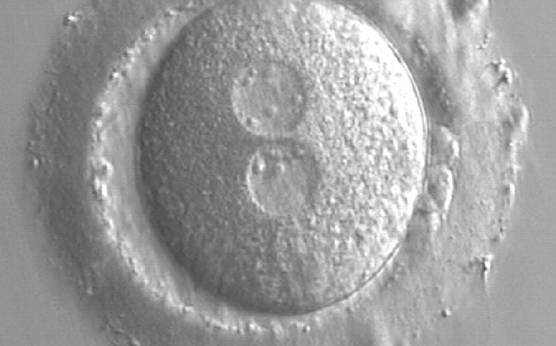
Figure 164
A zygote at 15 h post-IVF in which NPBs are scattered in one of the two PNs and aligned in the other (400× magnification). The ZP is thick and the PVS is enlarged. One of the two polar bodies is fragmented. It was transferred and implanted.
-
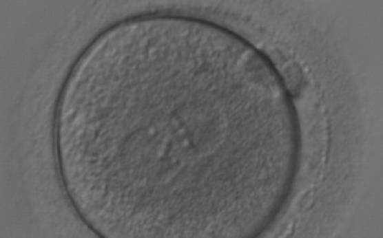
Figure 165
A zygote generated by ICSI with equal numbers of large-sized NPBs aligned at the PN junction (200× magnification). It was transferred but the outcome is unknown.
-
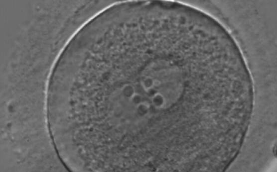
Figure 166
A zygote generated by ICSI with equal numbers of large-sized NPBs aligned at the PN junction (400× magnification). There is a halo in the cortical area; polar bodies are fragmented and the ZP appears brush-like. It was discarded due to subsequent abnormal development.
-
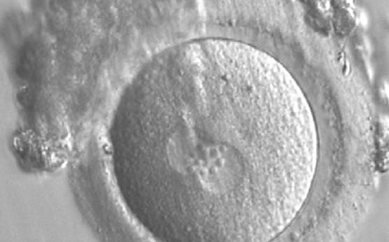
Figure 167
A zygote generated by IVF observed 17 h post-insemination (400× magnification). PNs have similar numbers of large-sized NPBs aligned at the PN junction. The PVS is slightly enlarged and the ZP is normal in size and surrounded by some granulosa cells. It was transferred but failed to implant.
-
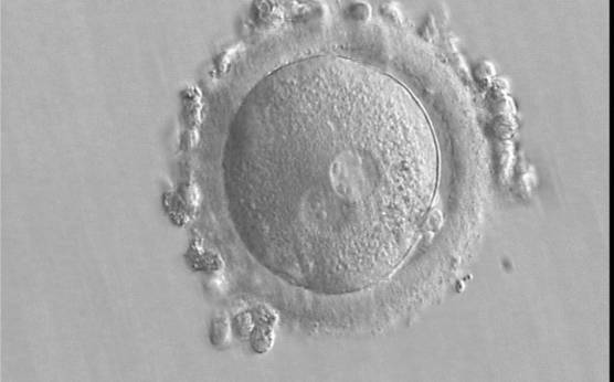
Figure 168
A zygote generated by IVF using frozen/thawed ejaculated sperm and observed 15 h post-insemination (400× magnification). Two PNs of approximately the same size are clearly visible in the cytoplasm. Peripheral granular cytoplasm can be seen. NPBs are scattered in both PNs. Both polar bodies are located at the 4 o'clock position. It was transferred and implanted.
-
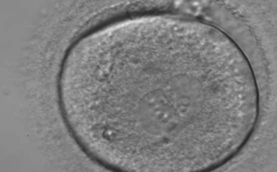
Figure 169
A zygote generated by ICSI showing different numbers of NPBs (400× magnification). The cytoplasm appears granular. It was transferred but failed to implant.
-
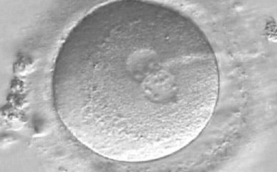
Figure 170
A zygote observed 16.5 h post-ICSI, showing unequal number and size of NPBs: medium-sized and scattered in one PN, larger-sized and aligned in the other (400× magnification). Polar bodies had been biopsied, and the slit opened mechanically in the ZP is evident at the 3 o'clock position. It was cryopreserved.
-
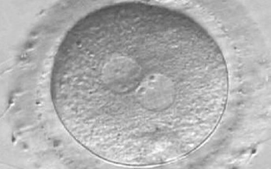
Figure 171
A zygote observed at 16 h post-IVF displaying different number and size of NPBs: small and scattered in one PN, large and aligned in the other (400× magnification). It was discarded because of developmental arrest.
-
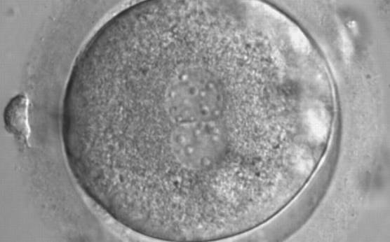
Figure 172
A zygote generated by ICSI with a different number and size of scattered NPBs (600× magnification). It was discarded because of subsequent abnormal development.
-
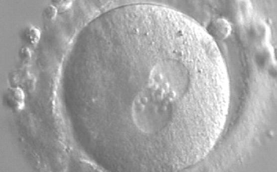
Figure 173
A zygote generated by ICSI showing equal number and size of NPBs, which are aligned at the PN junction (200× magnification). PNs are tangential to the plane of the polar bodies. It was transferred and resulted in a singleton pregnancy and delivery.
-
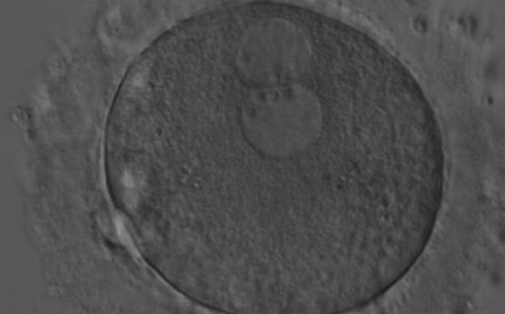
Figure 174
A zygote generated by ICSI with peripherally located PNs showing NPBs of similar size and number (200× magnification). Further development resulted in uneven cleavage and arrest on Day 3.
-
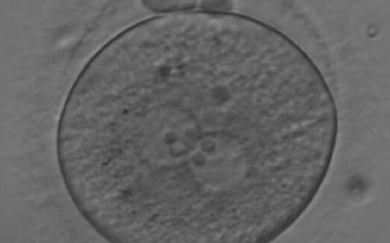
Figure 175
A zygote observed at 18 h after ICSI, with NPBs of equal size in both PNs and aligned at the PN junction (200× magnification). Polar bodies are intact and slightly larger than normal. The cytoplasm is granular with some inclusions. It was discarded due to subsequent abnormal development.
-
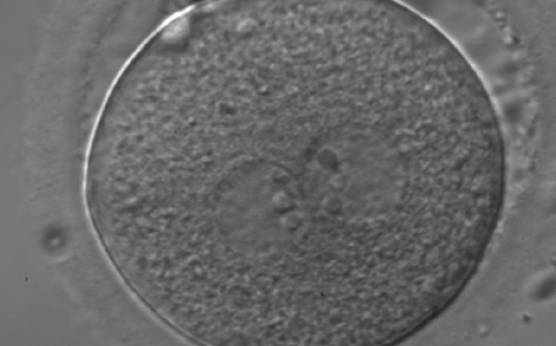
Figure 176
A zygote generated by ICSI showing equal number and size of NPBs, which are perfectly aligned at the PN junction (200× magnification). Polar bodies are highly fragmented. It was transferred but clinical outcome is unknown.
-
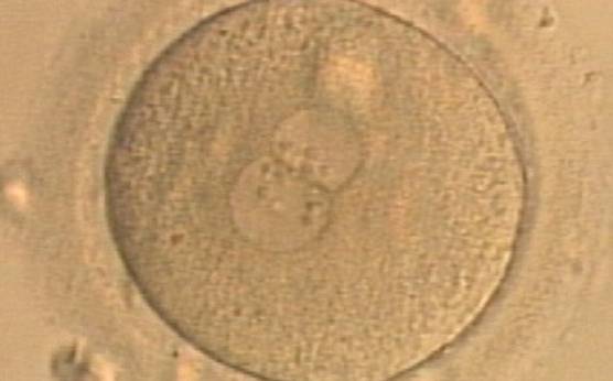
Figure 177
A zygote generated by ICSI displaying unequal number and size of NPBs between the PNs (400× magnification). It was transferred but clinical outcome is unknown.
-
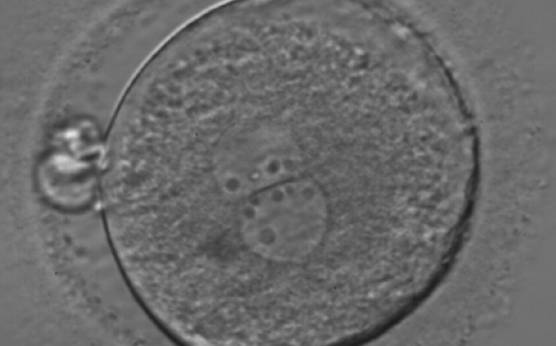
Figure 178
A zygote generated by ICSI showing an unequal number of NPBs (400× magnification). The NPBs differ in size within each PN. It was transferred but clinical outcome is unknown.
-
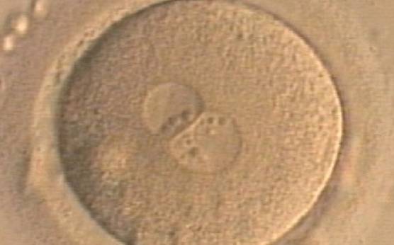
Figure 179
A zygote generated by ICSI displaying unequal number and size of NPBs between the PNs (400× magnification). NPBs are mainly aligned at the PN junction. It was transferred but clinical outcome is unknown.
-
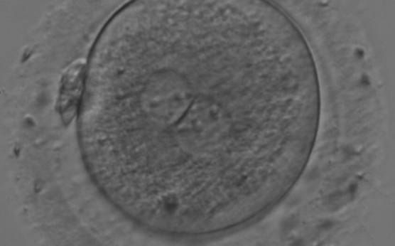
Figure 180
A zygote generated by IVF with large NPBs scattered with respect to the PN junction (200× magnification). NPBs display differences in sizes within each PN and are slightly larger in the PN on the right side. It was transferred and resulted in a singleton pregnancy with delivery.
-
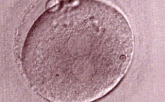
Figure 181
One out of three sibling zygotes (see also Figs 182 and 183) generated by ICSI using frozen/thawed epididymal sperm observed 18 h post-insemination. All three zygotes clearly show two distinct PNs with well-defined membranes and without any NPBs. All three were transferred and two healthy baby boys were delivered.
-
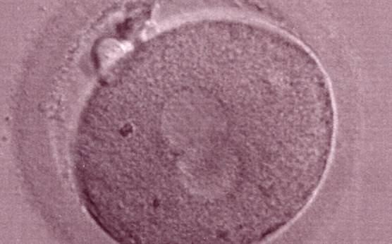
Figure 182
The second out of three sibling zygotes (see also Figs 181 and 183) generated by ICSI using frozen/thawed epididymal sperm. All three zygotes showed refractile bodies in the cytoplasm.
-
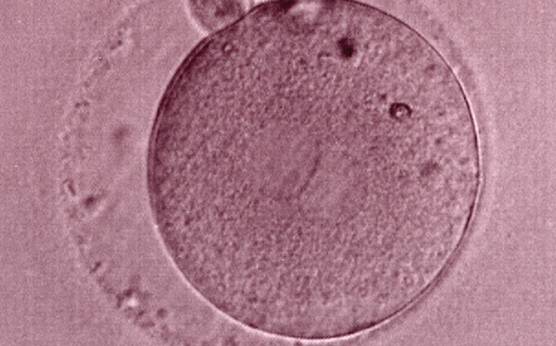
Figure 183
The third of three sibling zygotes (see also Figs 181 and 182) generated by ICSI using frozen/thawed epididymal sperm. Beside the absence of NPBs and the presence of refractile bodies, this zygote has a large perivitelline space.
-
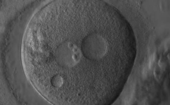
Figure 184
A zygote generated by ICSI showing what looks like two distinct PNs with distinct membranes and the absence of NPBs in one of the PNs (400× magnification). One small vacuole is visible under the left PN. Two highly fragmented polar bodies are present at 9 o'clock. Despite the presence of 2 polar bodies after ICSI, it is possible that this is a 1PN zygote, and that the structure to the right is a vacuole (compare with Fig. 209). It was not transferred.
-
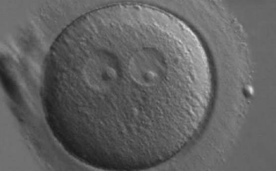
Figure 185
A zygote generated by ICSI showing two ‘bull's eye’ PNs, each having a single large NPB (200× magnification). The PNs are slightly separated and are not as yet juxtaposed. A clear cortical region is evident in the cytoplasm. It was discarded.
-
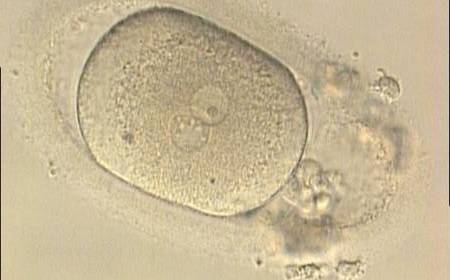
Figure 186
Oval-shaped zygote generated by ICSI, showing a single NPB in one of the two PNs (‘bull's eye’) and small, scattered NPBs in the other (200× magnification). The PVS is quite large and the polar bodies are highly fragmented. It was discarded.
-
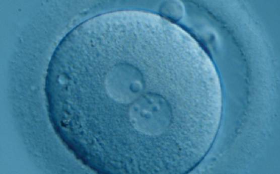
Figure 187
Zygote generated by ICSI with one ‘bull's eye’ NPB in one PN and small, different-sized NPBs in the other (400× magnification). It was transferred but clinical outcome is unknown.
-
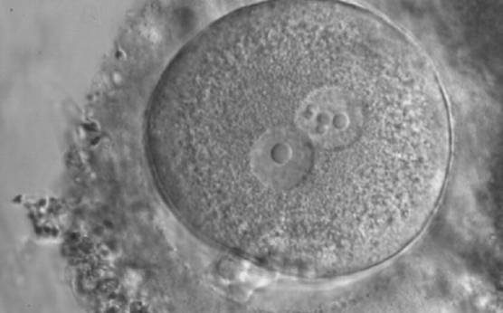
Figure 188
Zygote generated by ICSI displaying two centrally positioned PNs: in one PN there is a single large NPB (‘bull's eye’), whereas the NPBs are smaller, different-sized and scattered in the other (600× magnification). It was discarded.
-
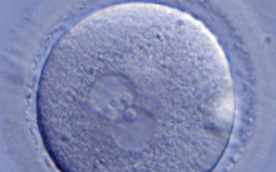
Figure 189
A zygote generated by ICSI with one ‘bull's eye’ PN (400× magnification). The NPBs from each PN are aligned at the PN junction. It was transferred but clinical outcome is unknown.