The Zygote
Cytoplasmic morphology assessment
Cytoplasmic morphology assessment
-
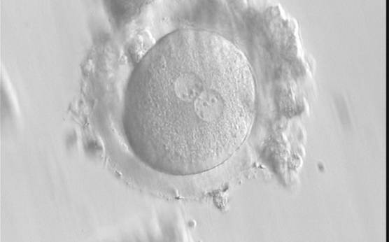
Figure 190
A zygote generated by IVF with frozen/thawed ejaculated sperm and observed at 16 h post-insemination showing normal cytoplasmic morphology (400× magnification). NPBs are obviously different in the two PNs. It was transferred but failed to implant.
-
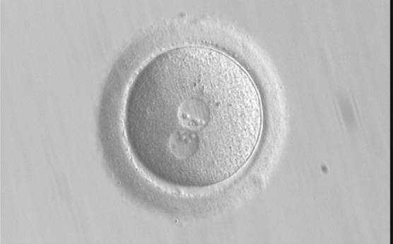
Figure 191
A zygote generated by ICSI, which subsequently underwent polar body biopsy, shows normal cytoplasmic morphology (400× magnification). It was discarded due to aneuploidy.
-
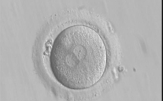
Figure 192
A zygote generated by IVF with frozen/thawed ejaculated sperm and observed at 16 h post-insemination showing normal cytoplasmic morphology with an evident clear cortical zone, the halo, in the cytoplasm (400× magnification). NPBs are scattered and different-sized, polar bodies are fragmented. It was cryopreserved.
-
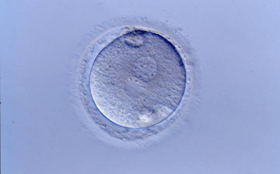
Figure 193
A zygote generated by ICSI showing normal cytoplasmic morphology, a thin ZP and small debris in an enlarged PVS (400× magnification). It was transferred but clinical outcome is unknown.
-
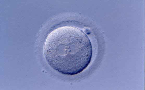
Figure 194
A zygote generated by ICSI showing normal cytoplasmic morphology except for a refractile body visible at the 3 o'clock position in this view (400× magnification). It was transferred but clinical outcome is unknown.
-
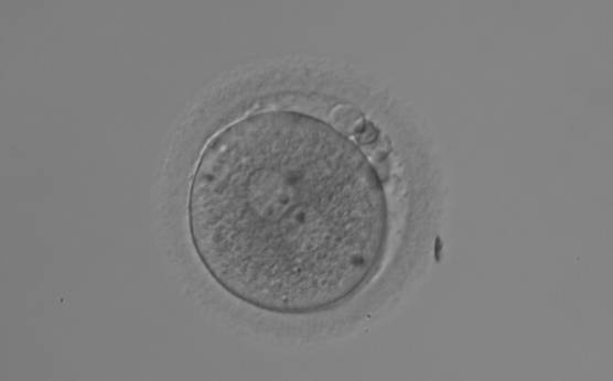
Figure 195
A zygote generated by ICSI displaying heterogeneous, granular cytoplasm (200× magnification). NPBs are large-sized and polar bodies are fragmented. It was discarded due to poor subsequent development.
-
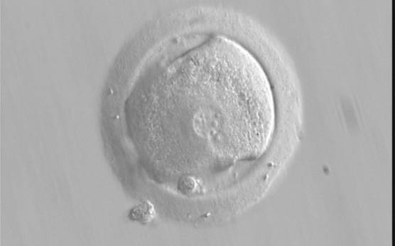
Figure 196
A zygote generated by IVF using frozen/thawed ejaculated sperm and observed at 17 h post-insemination showing an irregular oolemma and dysmorphic granular cytoplasm (400× magnification). PNs are different in size and peripherally located. NPBs differ in size and number between PNs. It was transferred but failed to implant.
-
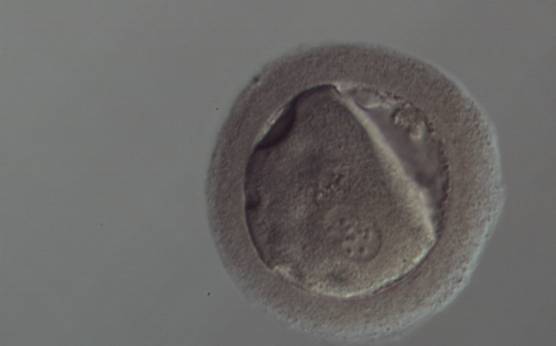
Figure 197
A zygote generated by ICSI with peripheral PNs (150× magnification). The oolemma is irregular and the cytoplasm is dysmorphic and granular. The ZP is thick and dark. It was discarded.
-
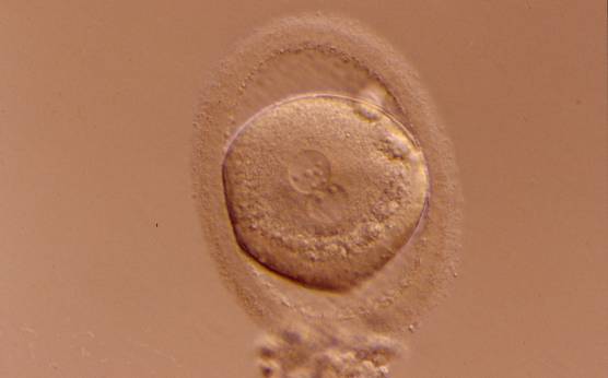
Figure 198
A zygote generated by ICSI displaying granular cytoplasm, especially in the area immediately adjacent to the clear cortical zone (400× magnification). There is an enlarged PVS and an ovoid ZP. NPBs differ in number and size. It was transferred but clinical outcome is unknown.
-
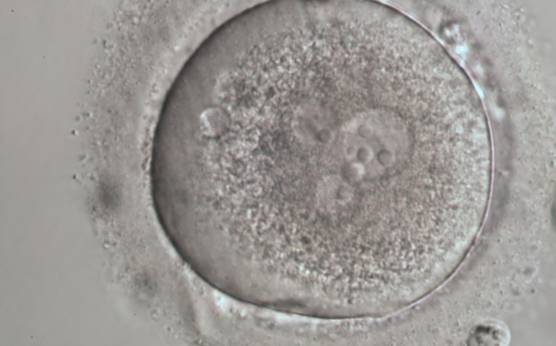
Figure 199
A zygote generated by ICSI with four PNs (possibly a result of fragmentation of an originally normal-sized PN), displaying very granular cytoplasm and a clear cortical zone (600× magnification). It was discarded.
-
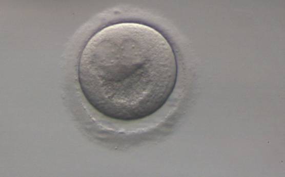
Figure 200
A zygote generated by ICSI using frozen/thawed ejaculated sperm (200× magnification). PNs are peripherally located with a large inclusion positioned directly below the PNs that is displaying a crater-like appearance as a consequence of severe organelle clustering. It was discarded.
-
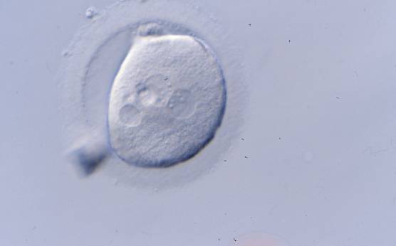
Figure 201
An irregularly shaped zygote generated by ICSI showing centrally positioned PNs and a medium-sized vacuole at the 8 o'clock position in the cytoplasm (400× magnification). It was transferred but clinical outcome is unknown.
-
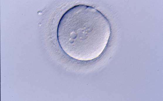
Figure 202
A zygote generated by ICSI with centrally positioned different-sized PNs, displaying two small vacuoles at the 9 o'clock position in the cytoplasm (400× magnification). It was transferred but clinical outcome is unknown.
-
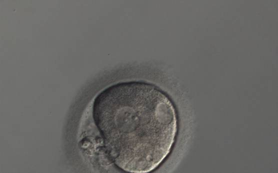
Figure 203
A zygote generated by ICSI with an irregular oolemma and a large PVS with possibly highly fragmented polar bodies (150× magnification). Different sized overlapping PNs are peripherally positioned and several vacuoles of different sizes are present at the 2, 5 and 6 o'clock position in the cytoplasm. It was discarded.
-
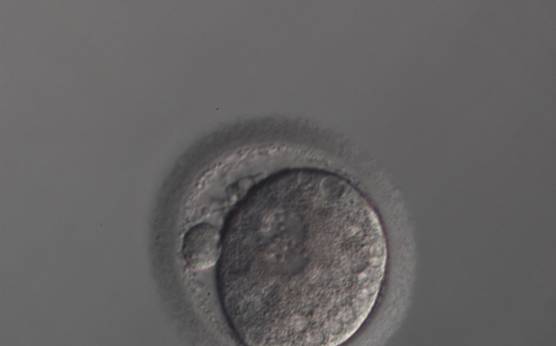
Figure 204
A zygote generated by ICSI with slightly overlapping peripherally positioned PNs and a large PVS with one large and one fragmented polar body (150× magnification). Many small vacuoles are present throughout the cytoplasm. It was discarded.
-
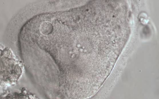
Figure 205
Severely dysmorphic zygote generated by IVF showing small PNs and an irregularly shaped ZP and oolemma and lack of a PVS (600× magnification). There is a small vacuole present at 10 o'clock with refractile bodies immediately adjacent. It was discarded.
-
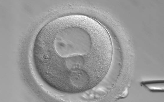
Figure 206
A zygote generated by ICSI showing peripherally positioned PNs and a very large vacuole at the 12 o′clock position in the cytoplasm (400× magnification). Polar bodies are fragmented and the ZP is of irregular thickness. It was discarded.
-
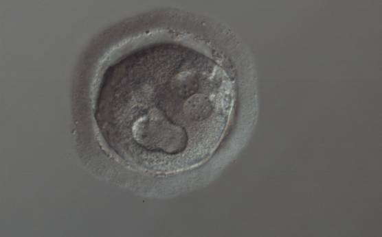
Figure 207
A zygote generated by ICSI showing cytoplasmic abnormalities (150× magnification). The PNs are juxtaposed and peripherally positioned with a large vacuole of irregular shape at the 6 o'clock position in the cytoplasm. The cytoplasm is granular and the ZP is thick and heterogeneous in appearance. It was discarded.
-
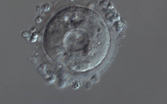
Figure 208
A zygote generated by ICSI showing severe cytoplasmic abnormalities (150× magnification). There is a large centralized vacuole with many small vacuoles surrounding it in a granular cytoplasm. It was discarded.
-
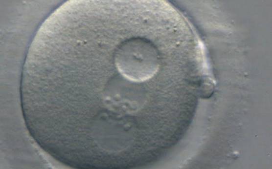
Figure 209
A zygote generated by ICSI showing peripherally located PNs with the same number and size of NPBs perfectly aligned at the PN junction (400× magnification). There is a large vacuole immediately adjacent to the two PNs that is almost the same size as the PNs. It was discarded.
-
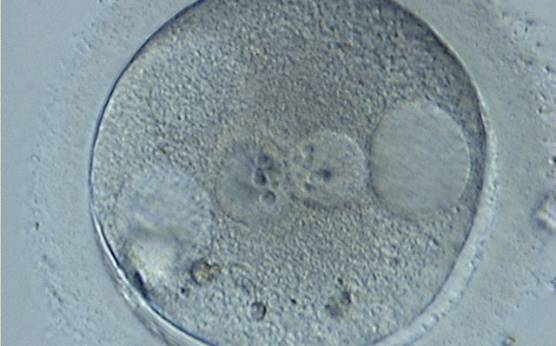
Figure 210
A zygote generated by ICSI showing centrally positioned PNs and two large vacuoles immediately adjacent to each of the PNs at the 3 and 9 o'clock positions in the cytoplasm (400× magnification). There are also refractile bodies present at the 6–7 o'clock positions and an area of clustering at 11 o'clock. It was discarded.