Blastocyst
Degree of expansion
Degree of expansion
-
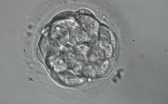
Figure 303
A very early blastocyst with a small cavity appearing centrally. The blastocyst was transferred but the outcome is unknown.
-
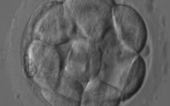
Figure 304
An early blastocyst with a cavity occupying <50% of the volume of the embryo. Note the early formation of the outer TE cells that are beginning to be flattened against the zona pellucida (ZP). The blastocyst was transferred and resulted in an ectopic pregnancy.
-
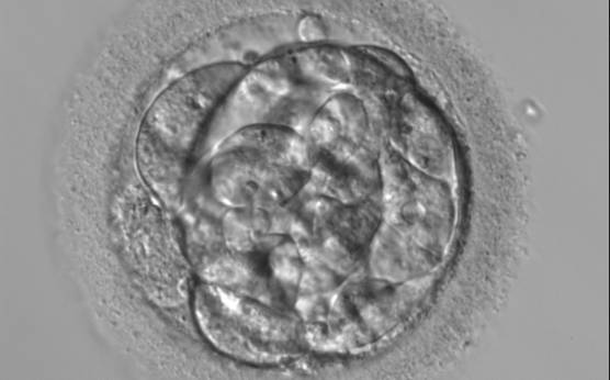
Figure 305
A very early blastocyst with a small cavity appearing centrally and can be seen most obviously at the 12 o'clock position in this view. Note the cellular debris that is not incorporated into the early blastocyst formation that has been sequestered to the perivitelline space (PVS).
-
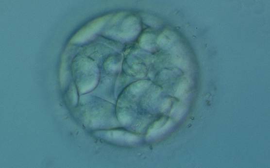
Figure 306
An early blastocyst with a cavity occupying <50% of the volume of the embryo. Note the flattened squamous-like trophectoderm (TE) cells lining the left half of the cavity in this view. The blastocyst was transferred but failed to implant.
-
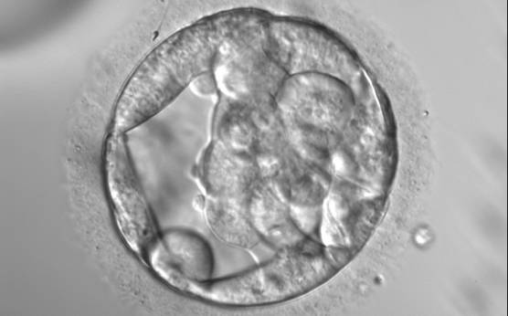
Figure 307
An early blastocyst with a cavity occupying almost 50% of the volume of the embryo. Note the large flattened TE cells lining the initial blastocoel cavity and the single spermatozoon embedded in the ZP at the 11 o'clock position in this view. The blastocyst was transferred but the outcome is unknown.
-
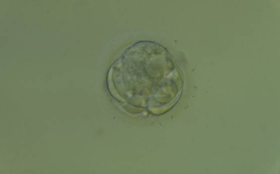
Figure 308
An early blastocyst with a cavity beginning to be formed centrally. Note the early formation of the flattened TE cells which at this stage are large, with two cells stretching to line the cavity from the 9 o'clock to the 3 o'clock position in this view. The blastocyst was transferred but failed to implant.
-
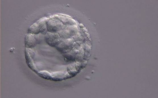
Figure 309
An early blastocyst with the cavity clearly visible and occupying half the volume of the embryo. The blastocyst was transferred but the outcome is unknown.
-
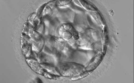
Figure 310
An early blastocyst with the cavity occupying >50% of the volume of the embryo. The overall volume of the blastocyst remains unchanged with no thinning of the ZP. The large cellular debris does not take part in the blastocyst formation.
-
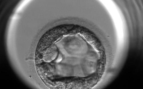
Figure 311
An early blastocyst with the cavity occupying >50% of the volume of the embryo. The TE cells are very large at this stage.
-
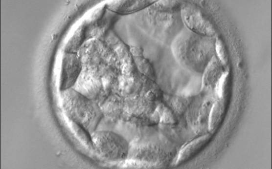
Figure 312
An early blastocyst with the cavity occupying >50% of the volume of the embryo. The overall volume of the blastocyst remains unchanged with no thinning of the ZP. The early ICM can be seen on the left half of the blastocyst in this view. The blastocyst was transferred and implanted.
-
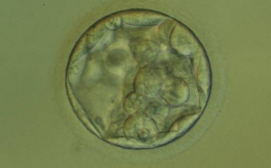
Figure 313
An early blastocyst with the cavity occupying >50% of the volume of the embryo. The overall volume of the blastocyst remains unchanged with no thinning of the ZP. The early ICM can be seen at the base of the blastocyst in this view. The blastocyst was transferred but failed to implant.
-
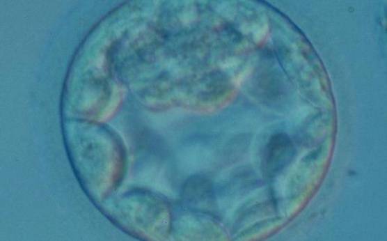
Figure 314
An early blastocyst with the cavity occupying >50% of the volume of the embryo. The overall volume of the blastocyst remains unchanged with no thinning of the ZP. The early ICM can be seen at the 12 o'clock position in this view. The blastocyst was transferred and resulted in a biochemical pregnancy.
-
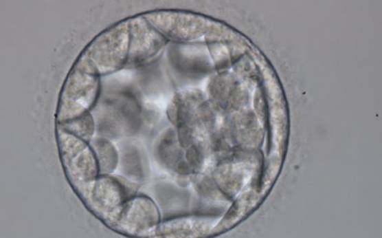
Figure 315
Blastocyst (Grade 3:2:1) showing a cavity occupying the total volume of the embryo. The ICM can be seen at the 3 o′clock position in this view and is loosely made up of only a few cells. The blastocyst was transferred but the outcome is unknown.
-
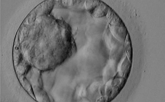
Figure 316
Blastocyst (Grade 3:1:1) showing a very large, mushroom-shaped ICM at the 10 o′clock position in this view. The ICM is made up of many cells that are tightly compacted. The blastocyst was transferred and resulted in the delivery of a healthy girl.
-
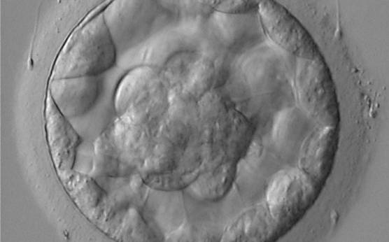
Figure 317
Blastocyst (Grade 3:1:1) showing a compact ICM at the base of the blastocyst in this view. The blastocyst was transferred and resulted in the delivery of a healthy boy.
-
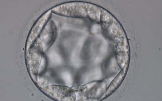
Figure 318
Blastocyst (Grade 3:3:2) with no clearly identifiable ICM and TE cells that in places are quite large and stretch over great distances to reach the next cell.
-
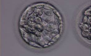
Figure 319
Blastocyst (Grade 3:1:1) with a dense ICM clearly visible at the 10 o′clock position in this view. The TE cells are variable in size but form a cohesive epithelium. The blastocyst was transferred but the outcome is unknown.
-
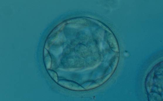
Figure 320
Blastocyst (Grade 3:1:1) with a dense, almost triangular, ICM clearly visible at the base of the blastocyst in this view. The blastocyst was transferred but failed to implant.
-
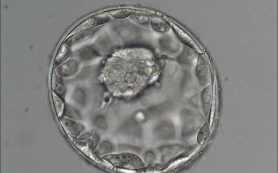
Figure 321
Good quality expanded blastocyst (Grade 4:1:1) with a large mushroom-shaped ICM. The blastocyst is now a greater volume than the original volume of the embryo and the ZP is thinned. There appears to be cytoplasmic strings extending from the ICM to the TE. The blastocyst was transferred and implanted.
-
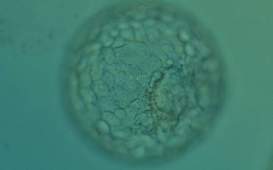
Figure 322
Expanded blastocyst (Grade 4:1:1) with an ICM clearly visible at the 4 o′clock position in this view. There are very many evenly sized cells making up a cohesive TE that surround the enlarged blastocoel cavity. The ZP is very thin. The blastocyst was transferred but the outcome is unknown.
-
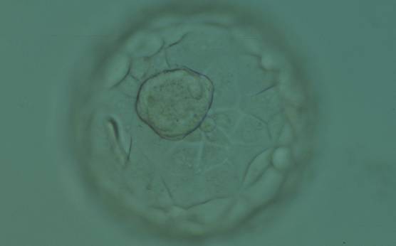
Figure 323
Expanded blastocyst (Grade 4:1:1) showing a large ICM at the base of the blastocyst in this view. The ICM is made up of many cells that are tightly compacted. The blastocyst volume is now larger than the original volume of the embryo causing the ZP to thin. The blastocyst was transferred but the outcome is unknown.
-
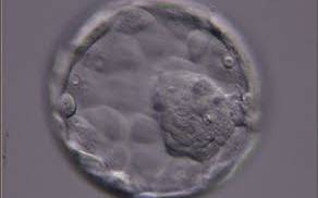
Figure 324
Expanded blastocyst (Grade 4:1:1) showing a large ICM at the 4 o′clock position in this view. The ICM is made up of many cells and is very compact. The blastocoel cavity is now larger than the original volume of the embryo and the ZP is very thin. The blastocyst was transferred but the outcome is unknown.
-
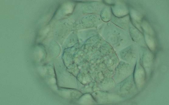
Figure 325
Expanded blastocyst (Grade 4:1:1) showing a large ICM at the base of the blastocyst in this view. There are very many TE cells forming a cohesive epithelium that lines the enlarged blastocoel cavity. The ZP is very thin. The blastocyst was transferred but failed to implant.
-
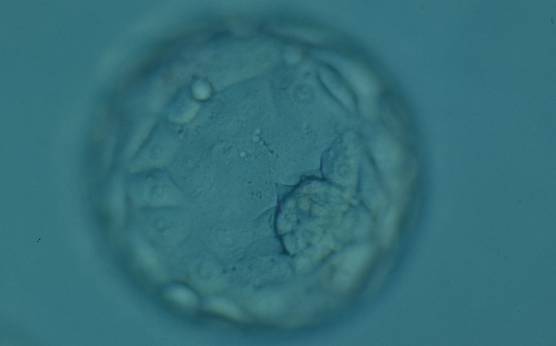
Figure 326
Expanded blastocyst (Grade 4:1:1) showing a compact ICM at the 4 o'clock position in this view. The blastocyst is now larger in volume than the original volume of the embryo causing the ZP to thin. The blastocyst was transferred but failed to implant.
-
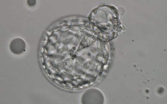
Figure 327
Hatching blastocyst (Grade 5:2:1) showing a small triangular ICM being drawn out along with the herniating TE cells at the 1 o'clock position in this view. There are very many TE cells of similar size lining the blastocoel cavity and the ZP is thinned. The blastocyst was transferred but the outcome is unknown.
-
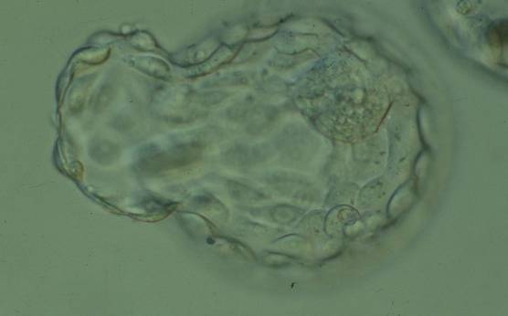
Figure 328
Hatching blastocyst (Grade 5:1:1) showing a large, compact ICM at the 1 o'clock position in this view. Approximately 25% of the blastocyst has herniated from a breach in the thinned ZP at the 8–10 o'clock positions in this view. The blastocyst was transferred but the outcome is unknown.
-
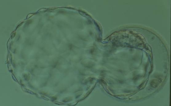
Figure 329
Hatching blastocyst (Grade 5:1:1) showing a large, compact, crescent-shaped ICM retained within the ZP at the 12 o'clock position in this view. There are very many TE cells and almost 75% of the blastocyst has herniated out through a breach in the ZP at the 8–10 o'clock positions in this view. The blastocyst was transferred but the outcome is unknown.
-
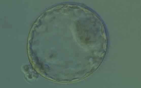
Figure 330
Hatching blastocyst (Grade 5:1:1) showing a large, compact ICM at the 2 o'clock position in this view. There are many TE cells of equivalent size lining the blastocoel cavity and several TE cells are herniating through a breach in the thinned ZP at the 8 o'clock position in this view. The blastocyst was transferred and implanted.
-
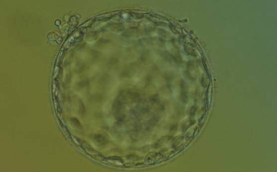
Figure 331
Hatching blastocyst (Grade 5:1:1) showing a large, compact ICM at the base of the blastocyst toward the 5 o'clock position and slightly out of focus in this view. There are very many TE cells of equivalent size making up a cohesive epithelium. Several TE cells have herniated out through a breach in the ZP at the 11 o'clock position in this view. The blastocyst was transferred and implanted.
-
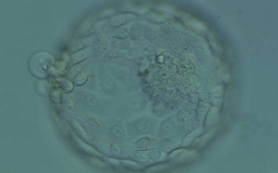
Figure 332
Hatching blastocyst (Grade 5:1:1) showing a large, compact and slightly granular ICM toward the 3 o'clock position in this view. There are very many TE cells of varying sizes making up a cohesive epithelium with several cells herniating out through a breach in the ZP at the 9 o'clock position in this view. The blastocyst was transferred but the outcome is unknown.
-
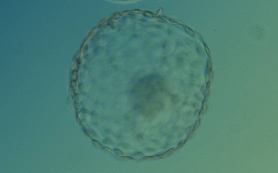
Figure 333
Hatched blastocyst (Grade 6:1:1) that is now completely free of the ZP. There is a large, compact ICM slightly out of focus at the base of the blastocyst toward the 4 o'clock position in this view. The blastocyst is now more than twice the size of the original expanded blastocyst. The blastocyst was transferred but failed to implant.
-
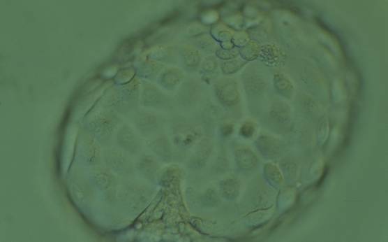
Figure 334
Hatched blastocyst (Grade 6:1:1) that is now completely free of the ZP showing a compact ICM at the 6 o'clock position in this view. The ICM appears to be connected or anchored to the TE by a broad triangular bridge. The TE cells vary in size but form a cohesive epithelium. The blastocyst is now more than twice the size of the original expanded blastocyst. The blastocyst was transferred but the outcome is unknown.
-
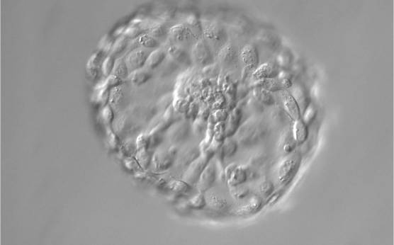
Figure 335
Hatched blastocyst (Grade 6:1:1) that is now completely free of the ZP. The ICM positioned centrally at the base of the blastocyst appears to be connected or anchored to the TE by several bridges. There are very many TE cells of similar size forming a cohesive epithelium. The blastocyst is now more than twice the size of the original expanded blastocyst.
-
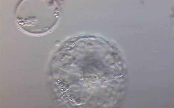
Figure 336
Hatched blastocyst (Grade 6:1:1) that is now completely free of the ZP which can be seen in the same view. Some cellular debris remains behind in the empty ZP. The ICM is large and compact at the base of the blastocyst and there are many TE cells making up a cohesive epithelium. In this view, it is possible to clearly see the increase in diameter of the blastocyst from the diameter of the original embryo which was accommodated within the ZP.
-
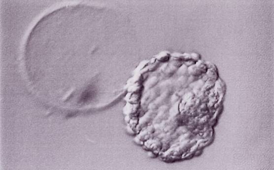
Figure 337
Hatched blastocyst (Grade 6:1:1) that is now completely free of the ZP which can be seen in the same view. The breach in the ZP is large. The ICM and TE both have many cells and the blastocyst has collapsed slightly and appears more dense. The blastocyst was transferred but the outcome is unknown.
-
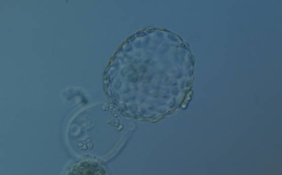
Figure 338
Hatched blastocyst (Grade 6:1:1) that is now only just free of the ZP which can also be seen in the low magnification view. The ICM is slightly out of focus in this view and is made up of many cells. The TE is similarly made up of many cells forming a cohesive epithelium. There is some cellular debris discarded in the ZP. The blastocyst was transferred but the outcome is unknown.
-
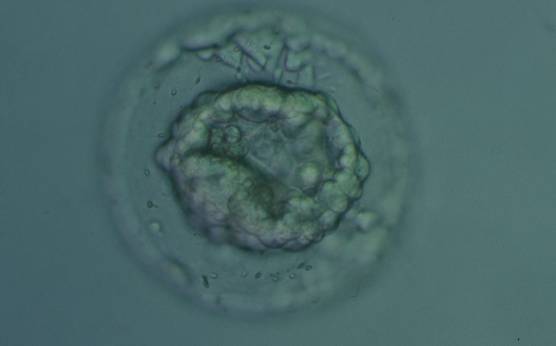
Figure 339
Collapsed blastocyst, which judging from the thickness of the ZP, is at least a Grade 3. The ICM and TE however cannot be accurately assessed. The blastocyst was transferred but the outcome is unknown.
-
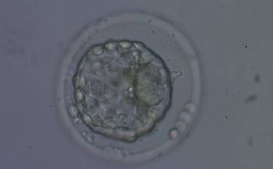
Figure 340
Collapsed blastocyst, which judging from the thickness of the ZP, is at least a Grade 3. In this view the ICM is clearly discernible at the 3 o'clock position and appears to be of Grade 1 quality. Similarly what can be seen of the TE cells appears to be Grade 1 in quality. There is some extraembryonic cellular debris in the PVS. The blastocyst was transferred but failed to implant.
-
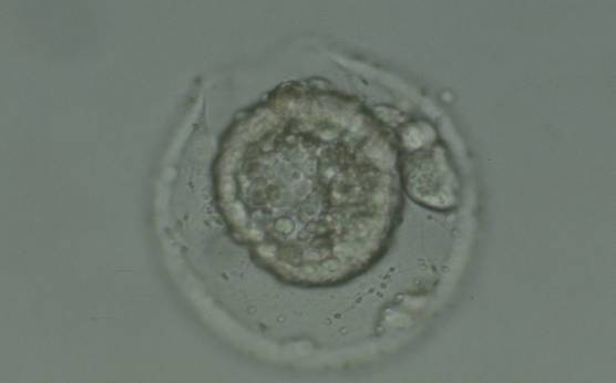
Figure 341
Collapsed hatching blastocyst (Grade 5) with the hatching site clearly visible as a breach in the ZP at the 11 o'clock position in this view. There is extraembryonic cellular debris both within the blastocoel cavity and external to the blastocyst in the PVS. Both the ICM and TE cannot be properly evaluated. The blastocyst was transferred but failed to implant.