Chapter 4
c. TE morphology
-
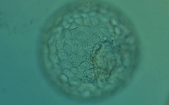
Figure 365
Expanded blastocyst (Grade 4:1:1) with many evenly sized cells making up a cohesive TE surrounding the enlarged blastocoel cavity. The ICM is clearly visible at the 4 o′clock position in this view. The blastocyst was transferred but the outcome is unknown.
-
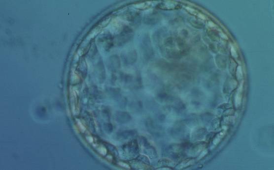
Figure 366
Early hatching blastocyst (Grade 5:1:1) with many cells making up a cohesive epithelium. The ICM is not clearly visible in this view. The blastocyst was transferred but failed to implant.
-
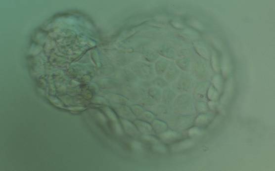
Figure 367
Hatching blastocyst (Grade 5:1:1) with many cells making up a cohesive TE. The ICM is herniating through a breach in the ZP at the 10 o′clock position in this view. The blastocyst was transferred but failed to implant.
-
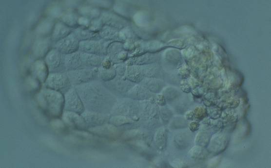
Figure 368
Hatching blastocyst (Grade 5:1:1) with many cells, some of variable size, making up a cohesive epithelium. The blastocyst was transferred but failed to implant.
-
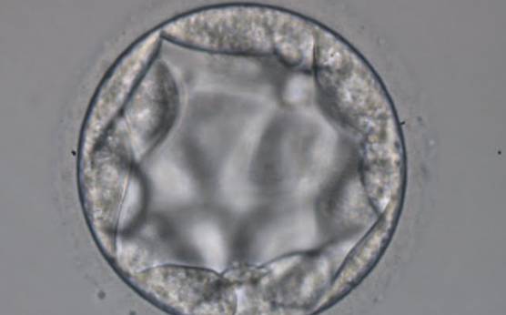
Figure 369
Blastocyst (Grade 3:3:2) with TE cells that in places are quite large and stretch over great distances to reach the next cell. No ICM can be identified.
-
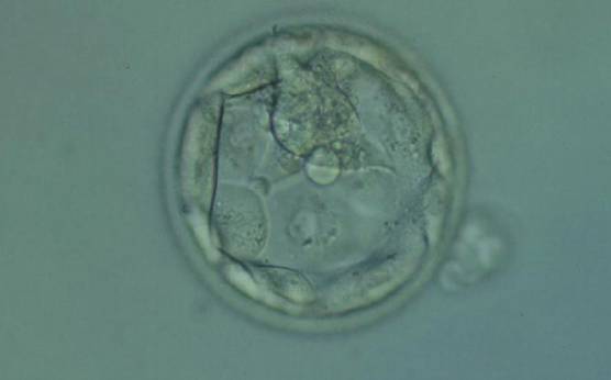
Figure 370
Hatching blastocyst (Grade 5.1.2) with few TE cells that form a loosely cohesive epithelium, and with a mushroom-shaped ICM at the 12 o'clock position in this view. The blastocyst was transferred but failed to implant.
-
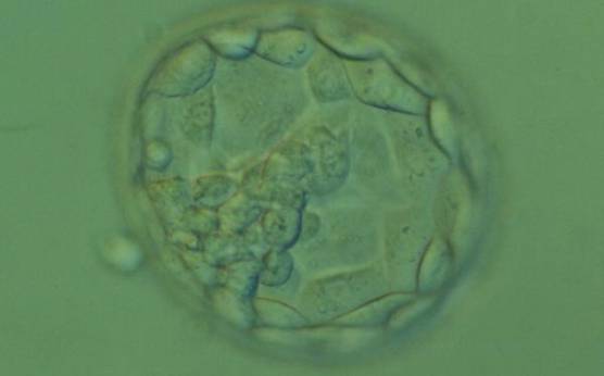
Figure 371
Hatching blastocyst (Grade 5.1.2). The TE cells vary in size with some cells quite large forming a loosely cohesive epithelium. A stellate ICM can be seen at the 8 o'clock position in this view. The blastocyst was transferred and failed to implant.
-
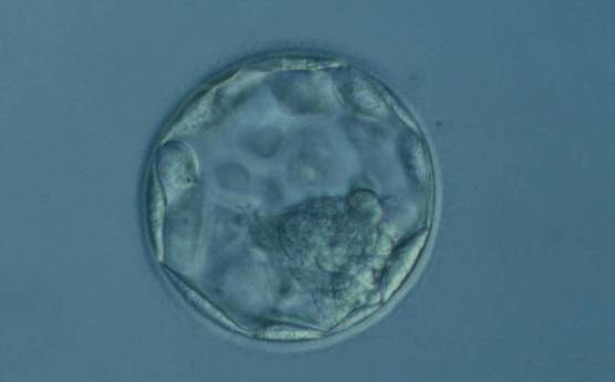
Figure 372
Expanded blastocyst (Grade 4:1:2). The TE cells vary in size with some cells quite large being particularly evident at the edge of the blastocyst where the cells stretch over some distance to reach their nearest neighbors. The blastocyst was transferred but the outcome is unknown.
-
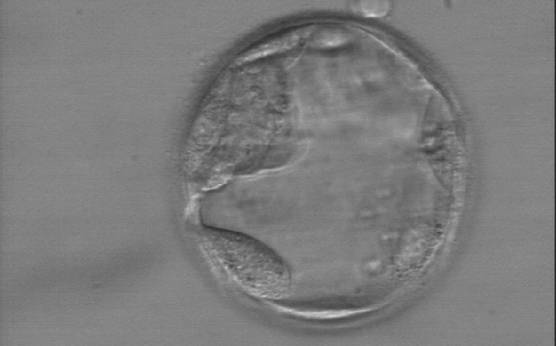
Figure 373
Early hatching blastocyst (Grade 5:1:3) with a very sparse TE that does not form a cohesive epithelium. The ICM is visible at the 10 o'clock position in this view.
-
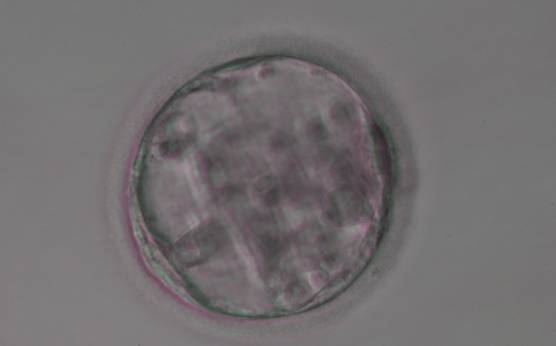
Figure 374
Blastocyst (Grade 3:3:3) with sparse TE that does not form a cohesive epithelium. The ICM is not clearly identifiable.
-
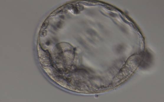
Figure 375
Expanded blastocyst (Grade 4:3:3) with sparse TE that does not form a cohesive epithelium. The ICM is hardly distinguishable despite the expansion of the blastocoel cavity.
-
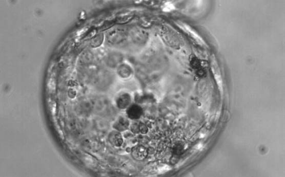
Figure 376
Hatching blastocyst (Grade 5:3:3). The TE varies in size and does not form a cohesive epithelium. Several loosely cohesive ICM cells can be seen at the 5 o'clock position in this view. There are several dark degenerate foci within the blastocyst.
-
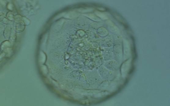
Figure 377
Expanded blastocyst (Grade 4:1:1). Note several dark granules within the majority of the TE cells. The ICM has several fragments. The blastocyst was transferred but failed to implant.
-
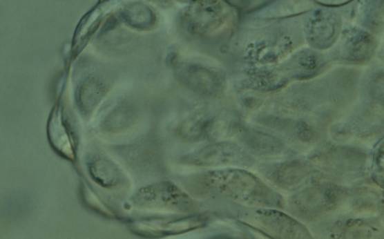
Figure 378
Hatching blastocyst (Grade 5:1:1) showing the herniating TE cells at high magnification. Many of the TE cells contain several dark granules. The blastocyst was transferred but the outcome is unknown.
-
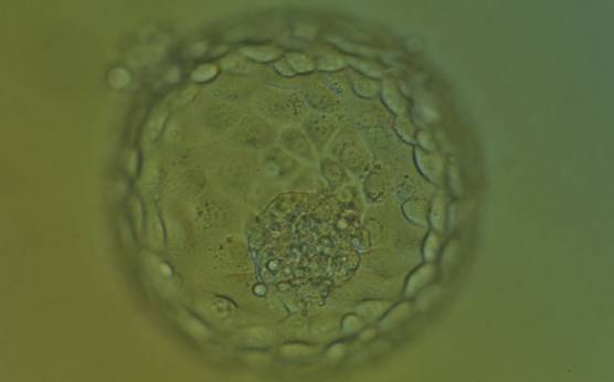
Figure 379
Hatching blastocyst (Grade 5:1:1) showing many of the TE cells contain dark granules. Several fragments are associated with a centrally positioned, compact ICM. The blastocyst was transferred and implanted.