B. Oocyte maturation stage
The removal of the cumulus–corona cell mass gives the unique opportunity to evaluate oocyte morphology prior to fertilization, and in particular, the nuclear maturation stage. Oocyte nuclear maturity, as assessed by light microscopy, is assumed to be at the MII stage when the PBI is visible in the PVS (Figs 10 and 11). The MII stage is characterized by the alignment of the homologous chromosomes on the spindle equatorial plate during metaphase of the second meiotic division. It is generally recognized that 85% of the retrieved oocytes following ovarian hyperstimulation display the PBI and are classified as MII, whereas 10% present an intracytoplasmic nucleus called the ‘germinal vesicle’ (GV; Figs 12–14), characteristic of prophase I of the first meiotic division. Approximately 5% of the oocytes have neither a visible GV nor PBI and these oocytes are generally classified as MI oocytes (Figs 15–17; Rienzi and Ubaldi, 2009). These oocytes may, however, be at the GV breakdown stage where the nuclear envelope has broken down but has not as yet progressed to true MI where the chromosomes are aligned on the metaphase plate in preparation for the completion of the first meiotic division.

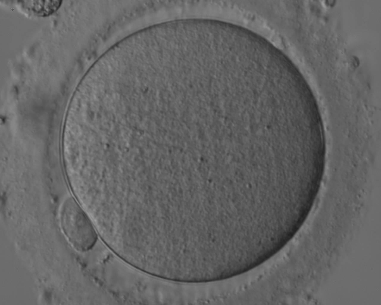
Figure 10
Denuded MII oocyte; an intact PBI is clearly visible in the PVS (400× magnification).
Figure 11
Denuded MII oocyte; the PBI is clearly visible in the narrow PVS (400× magnification).
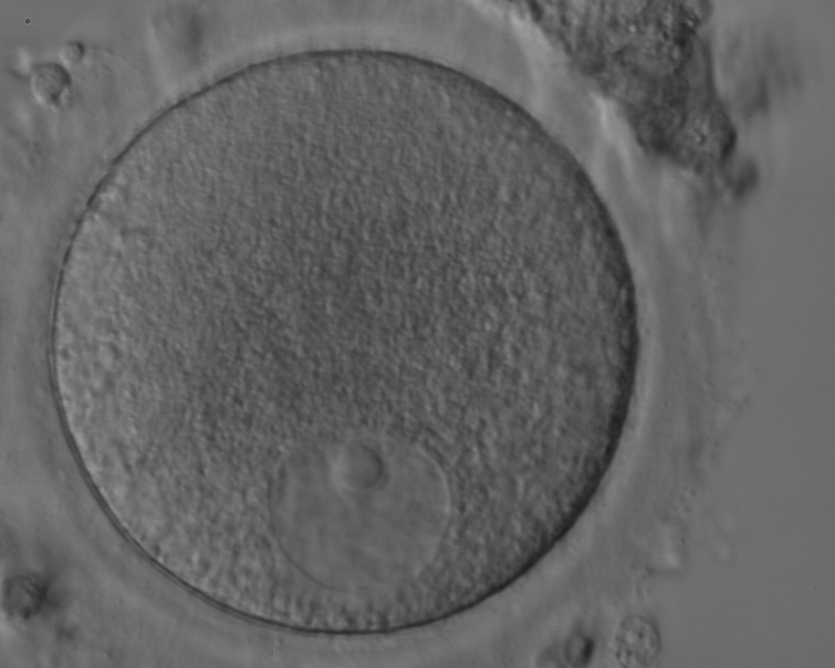

Figure 12
Denuded GV oocyte. A typical GV oocyte with an eccentrically placed nucleus and a prominent single nucleolus (400× magnification).
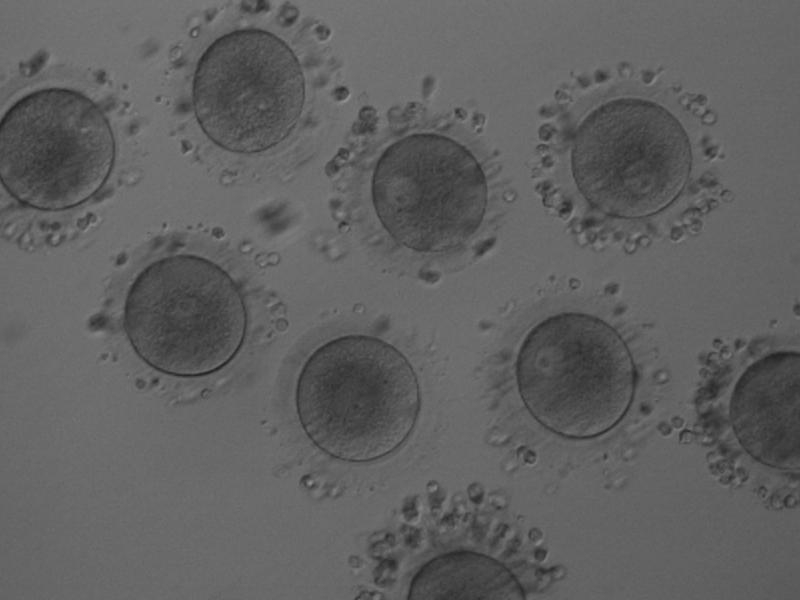

Figure 13
Denuded GV oocytes. Several GV with the organelles condensed centrally within the cytoplasm (200× magnification).
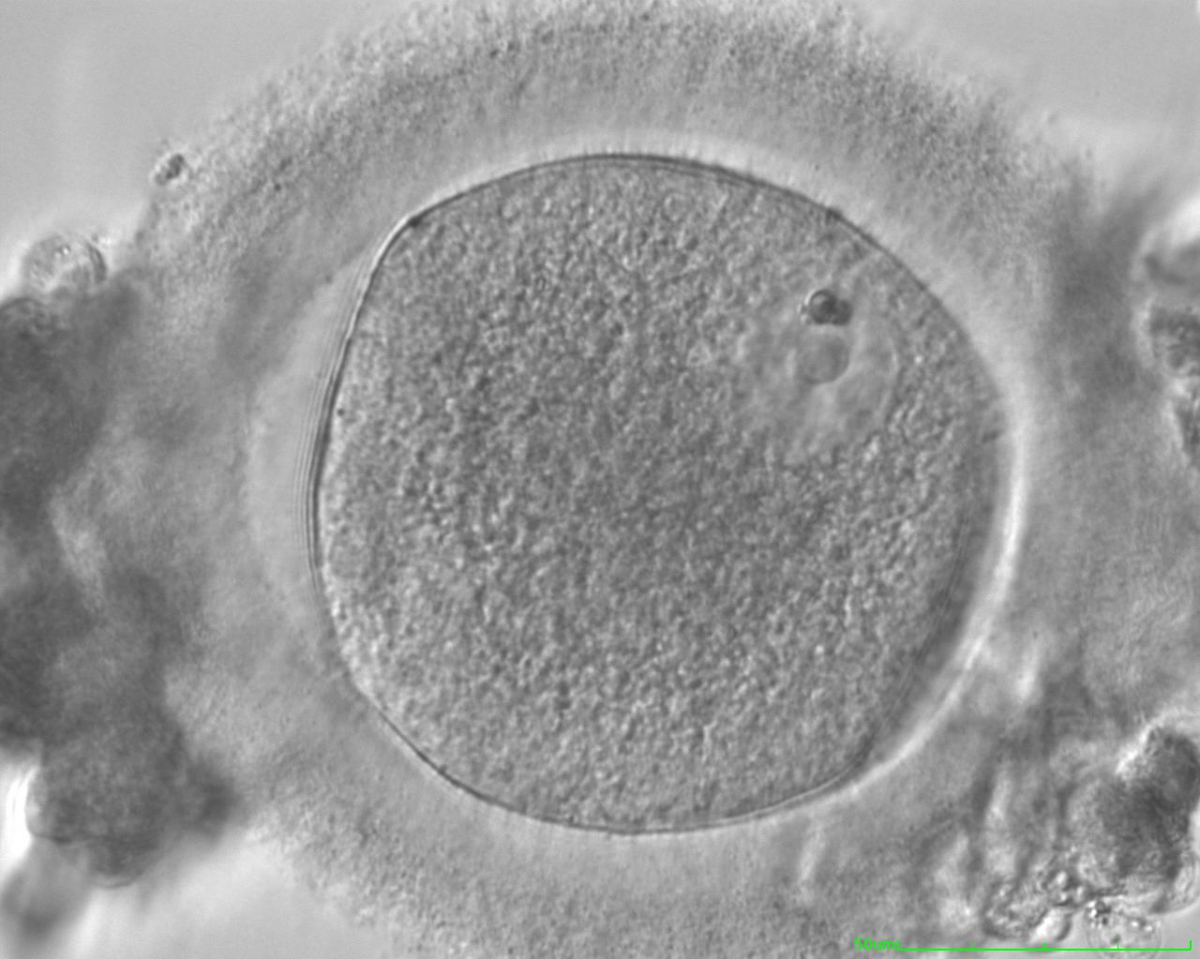
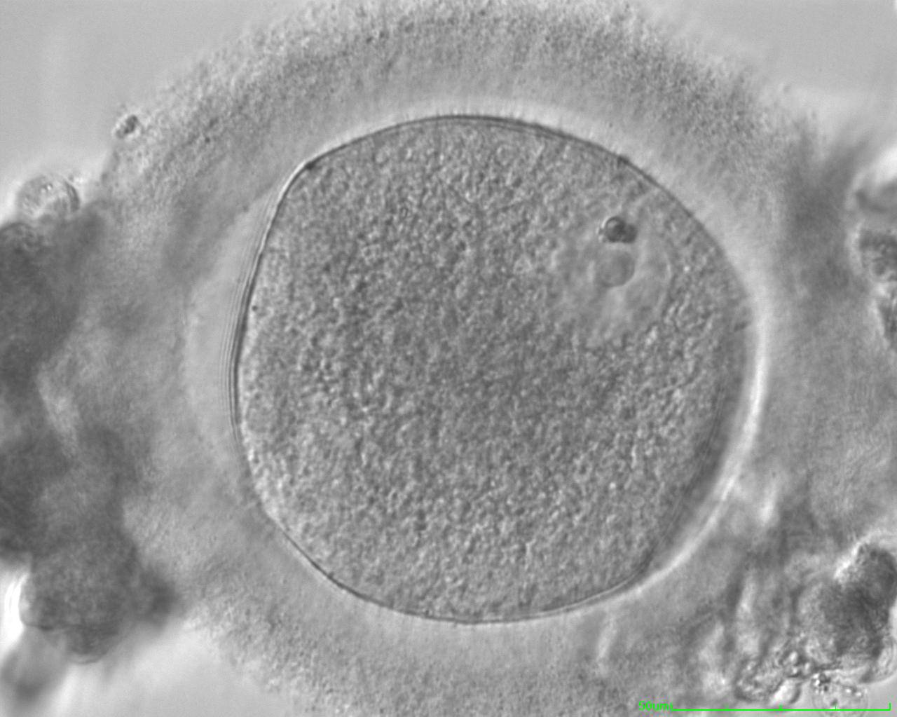
Figure 14
Denuded GV oocyte. A GV oocyte that is possibly approaching GVBD as the nuclear membrane is not distinct over its entirety (400× magnification).
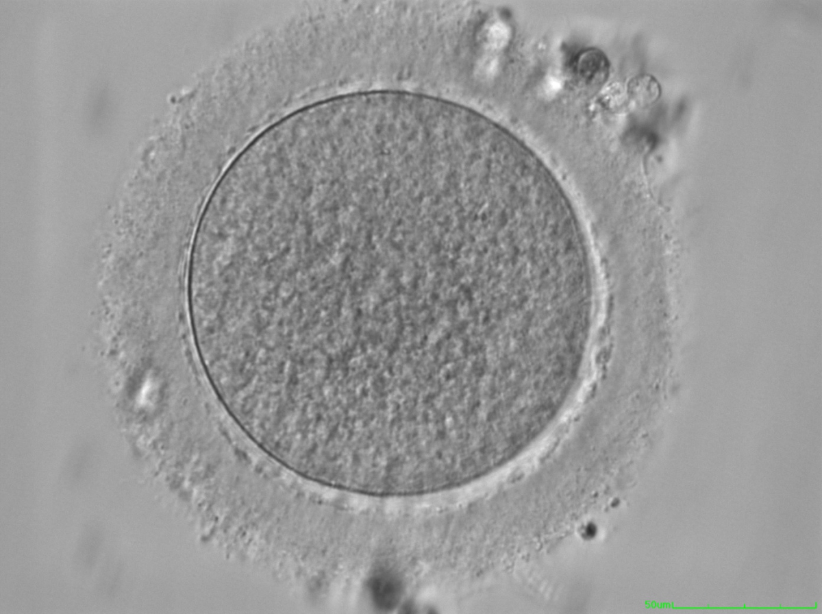
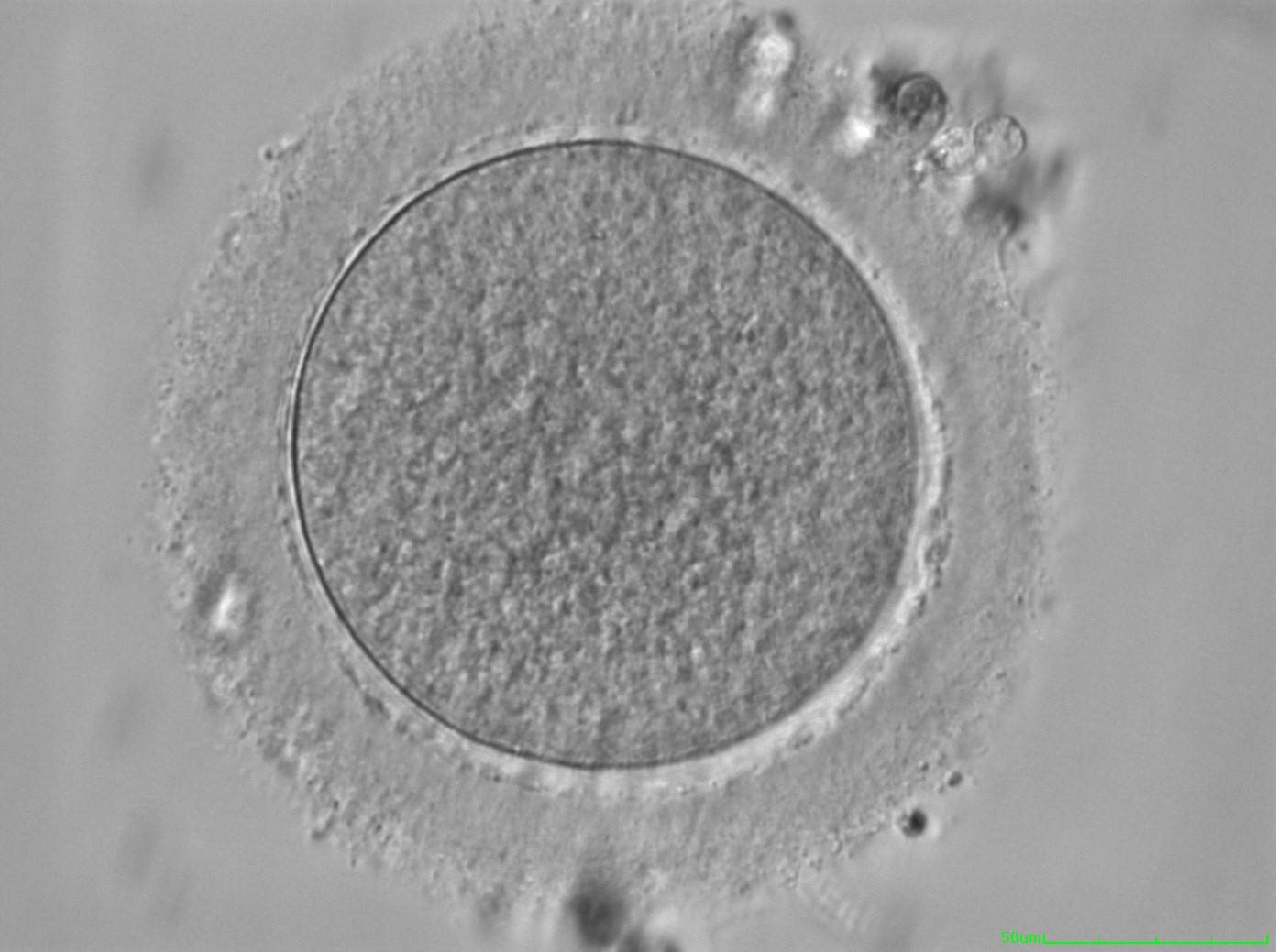
Figure 15
Denuded MI oocyte. This oocyte has no visible nucleus and has not as yet extruded the PBI (400× magnification). PVS is typically narrow.
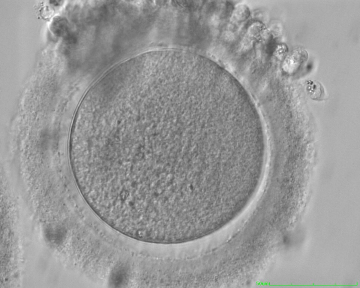
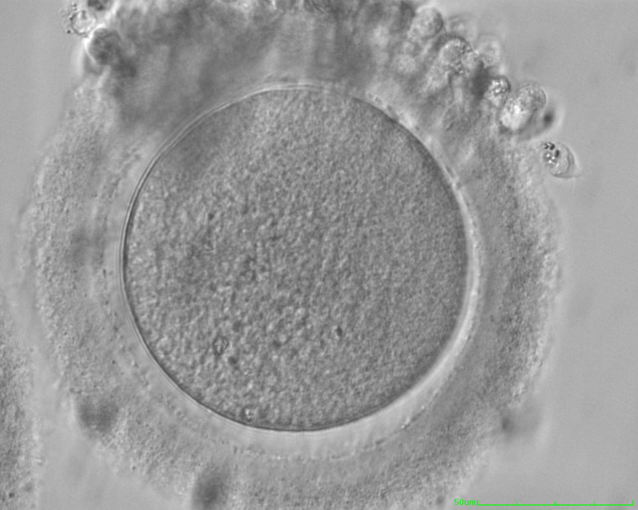
Figure 16
Denuded MI oocyte with no visible nucleus and no PBI (400× magnification). Some CCs are still tightly adhered to the ZP.

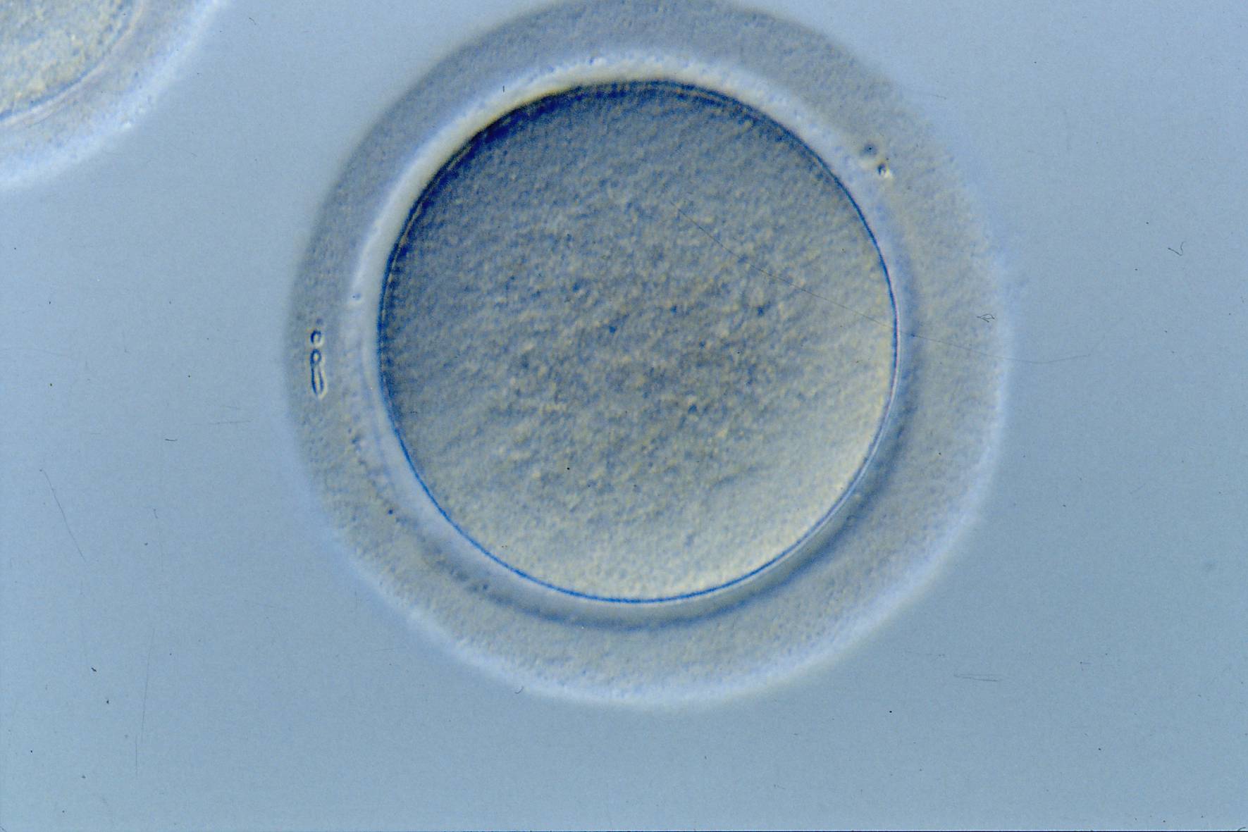
Figure 17
Denuded MI oocyte without a visible nucleus or an extruded PBI (400× magnification).
Additional information on oocyte nuclear status can be obtained with the use of polarized light microscopy combined with software for image processing for the non-invasive visualization of the MS and other oocyte birefringent structures. The MS is a microtubular structure involved in chromosome segregation, and therefore is crucial in the sequence of events leading to the correct completion of meiosis and subsequent fertilization. Parallel-aligned MS microtubules are birefringent and able to shift the plane of polarized light inducing a retardance; these properties enable the system to generate contrast and image the MS structure (Oldenbourg, 1999; Fig. 18). The presence of the MS gives more accurate information about the nuclear stage of the oocyte. In particular, some oocytes can be immature (at the stage of early telophase I) when observed with polarized light microscopy, despite the presence of PBI in the PVS. At this stage, in fact, there is continuity between the ooplasm of the oocyte and the forming PBI and the MS is interposed between the two separating cells (Figs 19–22). This condition normally has a duration of 75–90 min. The MS has been found to disappear in late telophase I (Fig. 23), reforming only 40–60 min later (Montag et al., 2011). However, it must be underlined that other factors, such as sub-optimal culture conditions, temperature fluctuations and chemical stress during manipulation, can contribute to MS disassembly (Rienzi and Ubaldi, 2009). Finally, the percentage of oocytes with detectable MS is also related to the time elapsed from HCG administration and is higher after 38 h (Cohen et al., 2004). In general, it is expected that at least 80% of oocytes recovered following ovarian hyperstimulation are MS positive when viewed by polarized light microscopy.

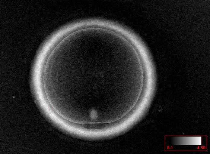
Figure 18
MII oocyte visualized using polarized light microscopy (400× magnification). The polar body is present at the 6 o'clock position in the PVS, and the MS of the second meiotic division is visible in the cytoplasm perfectly aligned to PB1 position. This is a fully mature MII oocyte.

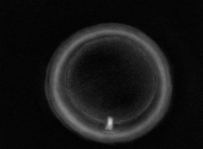
Figure 19
Telophase I oocyte visualized using polarized light microscopy (400× magnification). PB1 is present in the PVS; however, the MS can be seen between PB1 and the oocyte cytoplasm indicating that this oocyte is still completing the first meiotic division. This is not yet a fully mature MII oocyte.

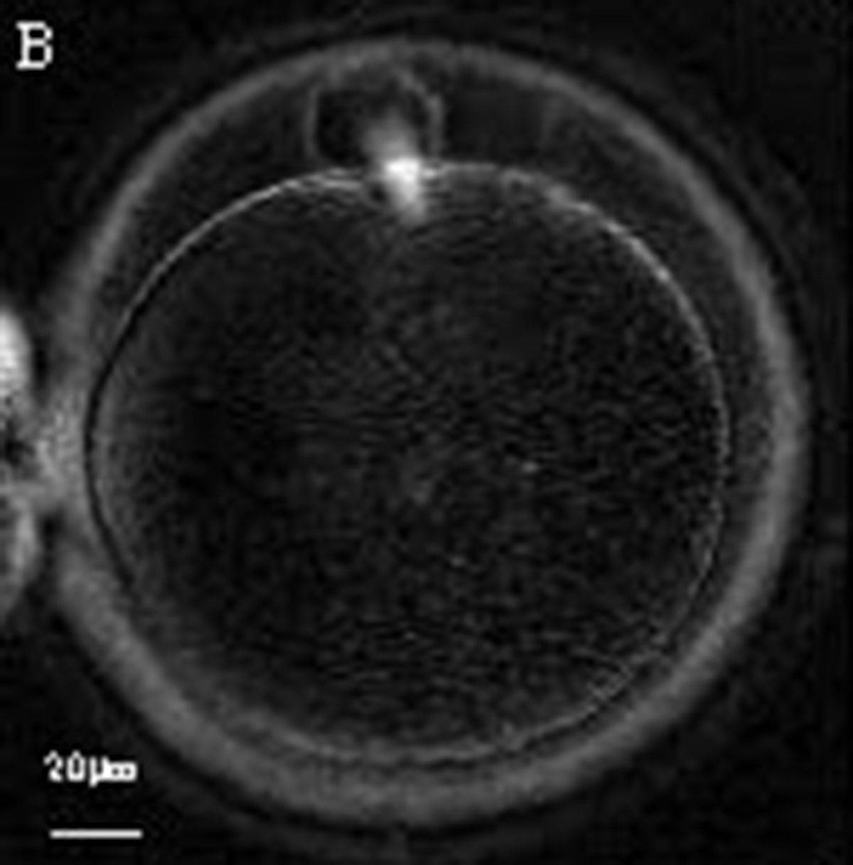
Figure 20
Telophase I oocyte visualized using polarized light microscopy (400× magnification). The MS can be seen between PB1 and the oocyte cytoplasm indicating that the first meiotic division is not yet completed.

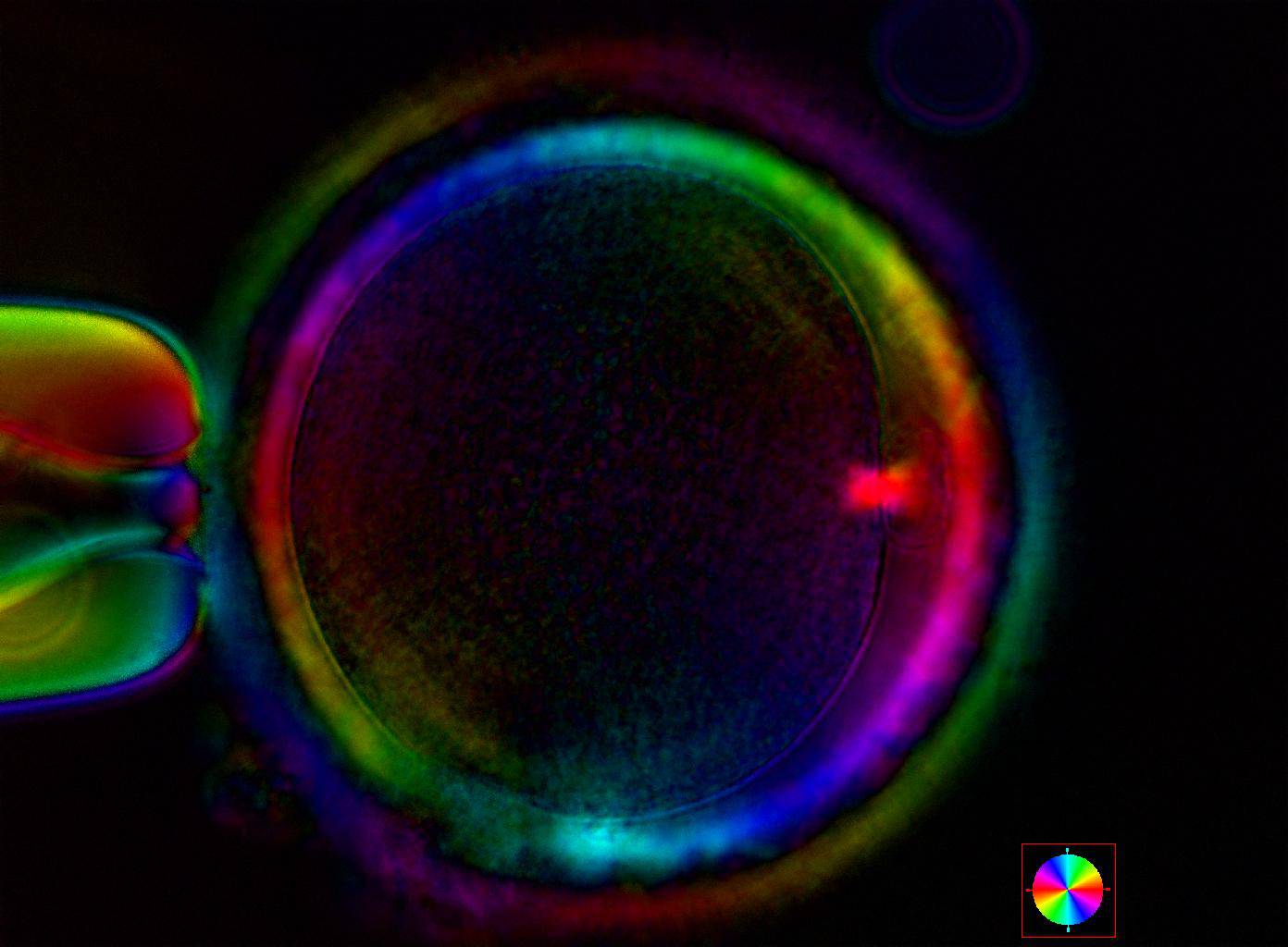
Figure 21
Telophase I oocyte visualized using polarized light microscopy (400× magnification). PB1 is present in the PVS at the 3 o'clock position; however, the MS is still bridging PB1 and the oocyte cytoplasm indicating that this oocyte is not yet a fully mature MII oocyte.
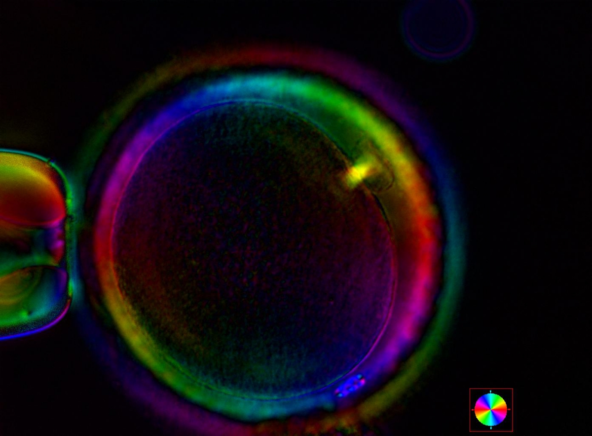
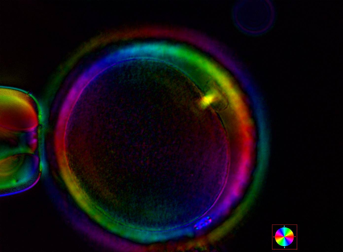
Figure 22
Telophase I oocyte visualized using polarized light microscopy (400× magnification). The MS can be seen between PB1 and the oocyte cytoplasm indicating that this oocyte is still completing the first meiotic division despite the extrusion of PB1 in the PVS.

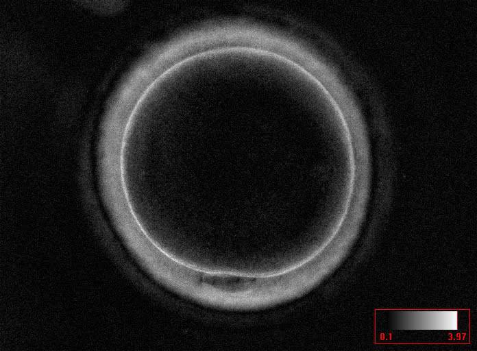
Figure 23
Interphase oocyte (between the first and second meiotic division; Prophase II) visualized using polarized light microscopy (400× magnification). PB1 is present in the PVS at the 6 o'clock position; however, the MS of the second meiotic division is not yet visible in the cytoplasm. This is not yet a fully mature MII oocyte.
Article references:
Cohen Y, Malcov M, Schwartz T, Mey-Raz N, Carmon A, Cohen T, Lessing JB, Amit A, Azem F. Spindle imaging: a new marker for optimal timing of ICSI? Hum Reprod 2004;19:649-654.
Abstract/FREE Full Text
Oldenbourg R. Polarized light microscopy of spindles. Methods Cell Biol 1999;61:175-208.
Medline | Web of Science | Google Scholar
Rienzi L, Ubaldi F. Oocyte retrieval and selection. In: Gardner DK, Weissman A, Howles CM, Shoham Z, editors. Textbook of Assisted Reproductive Technologies: Laboratory and Clinical Perspectives. 3rd edn. London, UK: Informa Healthcare; 2009. p. 5-101.
Google Scholar
Montag M, Köster M, Van der Ven K, van der Ven H. Gamete competence assessment by polarizing optics in assisted reproduction. Hum Reprod Update 2011;17:654-666.
Abstract/FREE Full Text
