E. Cytoplasmic anomalies
The cytoplasm of cleaving embryos is normally pale, and clear or finely granular in appearance (Hartshorne, 2000). Cytoplasmic anomalies, such as cytoplasmic granularity, cytoplasmic pitting and the presence of vacuoles, occur occasionally and can also be scored in the morphological assessment of Days 2 and 3 embryos. However, a possible predictive value of these features to embryo quality or implantation potential is unclear.
Cytoplasmic pitting (Figs 271 and 272) is characterized by the presence of numerous small pits with an approximate diameter of 1.5 µm on the surface of the cytoplasm (Biggers and Racowksy, 2002). Although cytoplasmic pitting in Day 3 embryos seems to be associated with improved blastocyst formation, the appearance of cytoplasmic granularity has no prognostic value to embryo quality (Rienzi et al., 2003) or to pregnancy (Desai et al., 2000). Other studies have shown that culture conditions may induce cytoplasmic pitting (Biggers and Racowsky, 2002; Ebner et al., 2005b) which in extreme cases may result in an increased risk of early loss of gestational sacs (Ebner et al., 2005b).

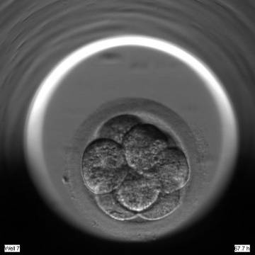
Figure 271
An 8-cell embryo with equally sized blastomeres showing cytoplasmic pitting. Numerous small pits are present on the surface of the cytoplasm.
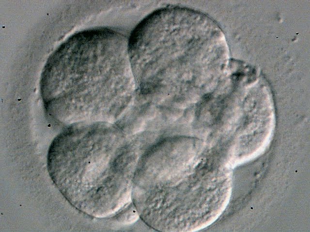
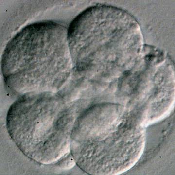
Figure 272
An 8-cell embryo with equally sized blastomeres showing cytoplasmic pitting. Numerous small pits are homogeneously distributed in the cytoplasm. The 5 blastomeres in focus are arranged in one spatial plane.
The cytoplasm of blastomeres may be excessively darkened with centralized granularity associated with a cortical halo, as cytoplasmic organelles retract toward the center of the blastomere (Fig. 273). It was suggested that these embryos have reduced implantation potential or are destined for degeneration (Veeck, 1999). Similarly, embryos with alternating areas of granularity and clear zones within the blastomeres are even more likely to degenerate (Fig. 274).

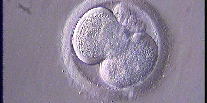
Figure 273
A 2-cell embryo with a clear halo in both blastomeres, characterized by centralized granularity associated with an absence of organelles in the peripheral cortex.
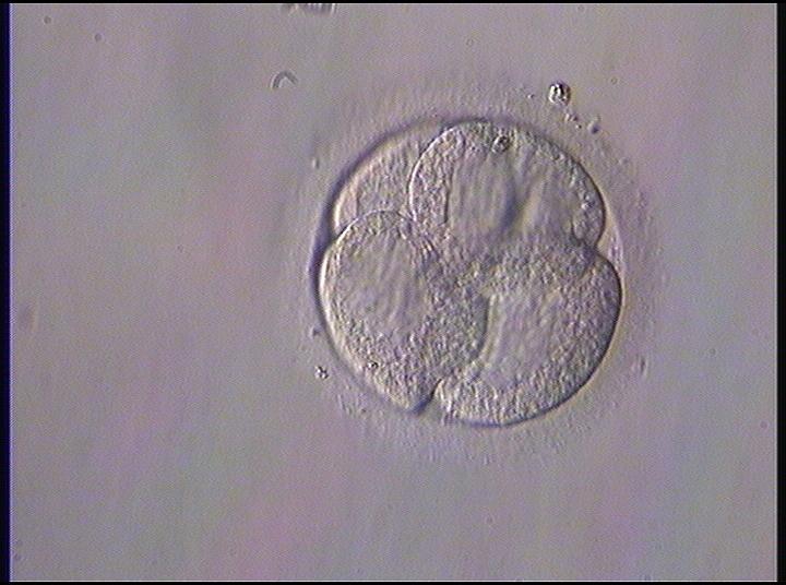
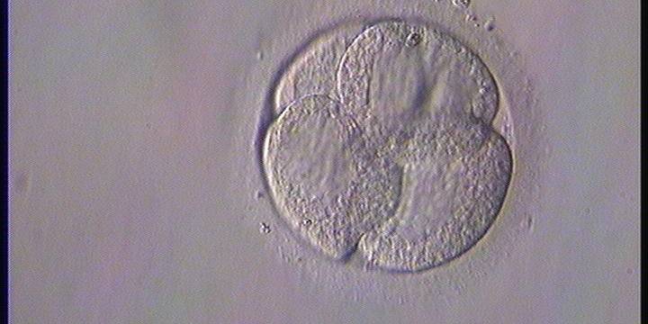
Figure 274
A 4-cell embryo on Day 2 with an abnormal distribution of organelles leading to differential granular and smooth zones inside each cell.
Cytoplasmic vacuolization is probably the most common cytoplasmic dysmorphism in human oocytes/embryos. Vacuoles vary in size and in number (Figs 275–280). They are membrane-bound cytoplasmic inclusions filled with fluid that are virtually identical with the perivitelline fluid (Van Blerkom, 1990). Whereas vacuoles have been well studied and described in human oocytes, very little is known about their incidence and role in developing embryos. Beside vacuoles visible at the time of oocyte collection and those created artificially by ICSI, vacuoles may also arise at the compaction stage (Ebner et al., 2005a). De novo formation of vacuoles on Day 4 is related to developmental arrest with a detrimental effect on blastocyst formation (Ebner et al., 2005a). It is believed that the occurrence of a few, small vacuoles (Figs 275 and 276) is not of importance (Alpha Scientists in Reproductive Medicine and ESHRE Special Interest Group of Embryology, 2011), but in cases of extensive vacuolization (Figs 277–280) it may be detrimental, mainly to spatial development, and the assessment should be added to the selection score.
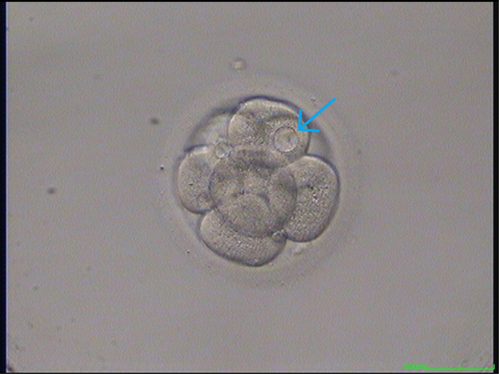
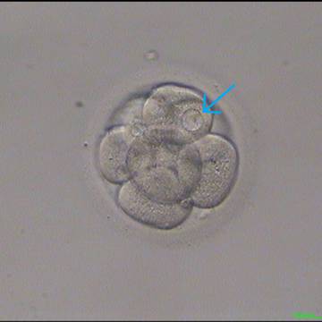
Figure 275
An 8-cell embryo with one blastomere showing a small vacuole (arrow).
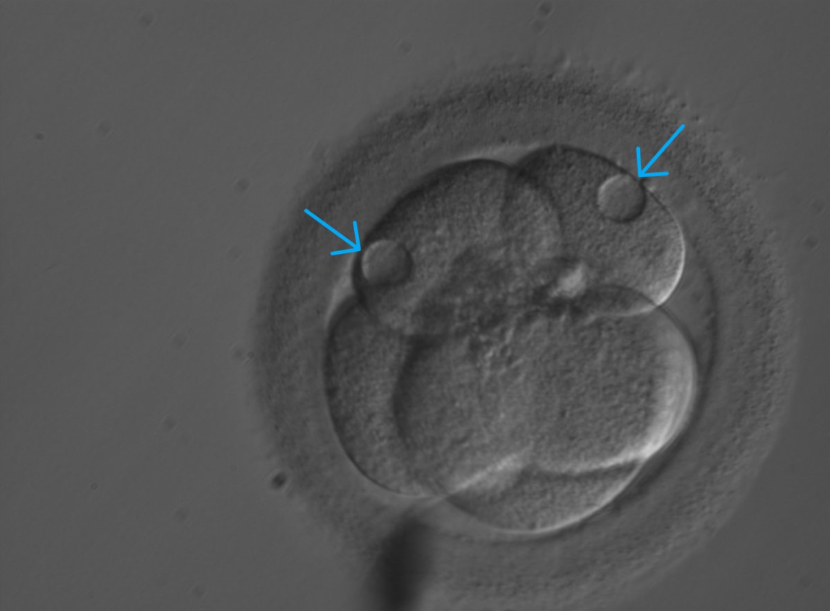
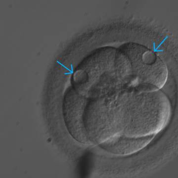
Figure 276
A 5-cell embryo with two small and three large blastomeres. There is a small vacuole in each of the two smaller blastomeres.
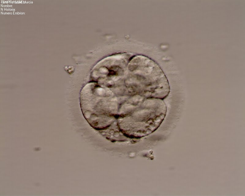
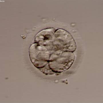
Figure 277
An embryo with abundant small vacuoles.
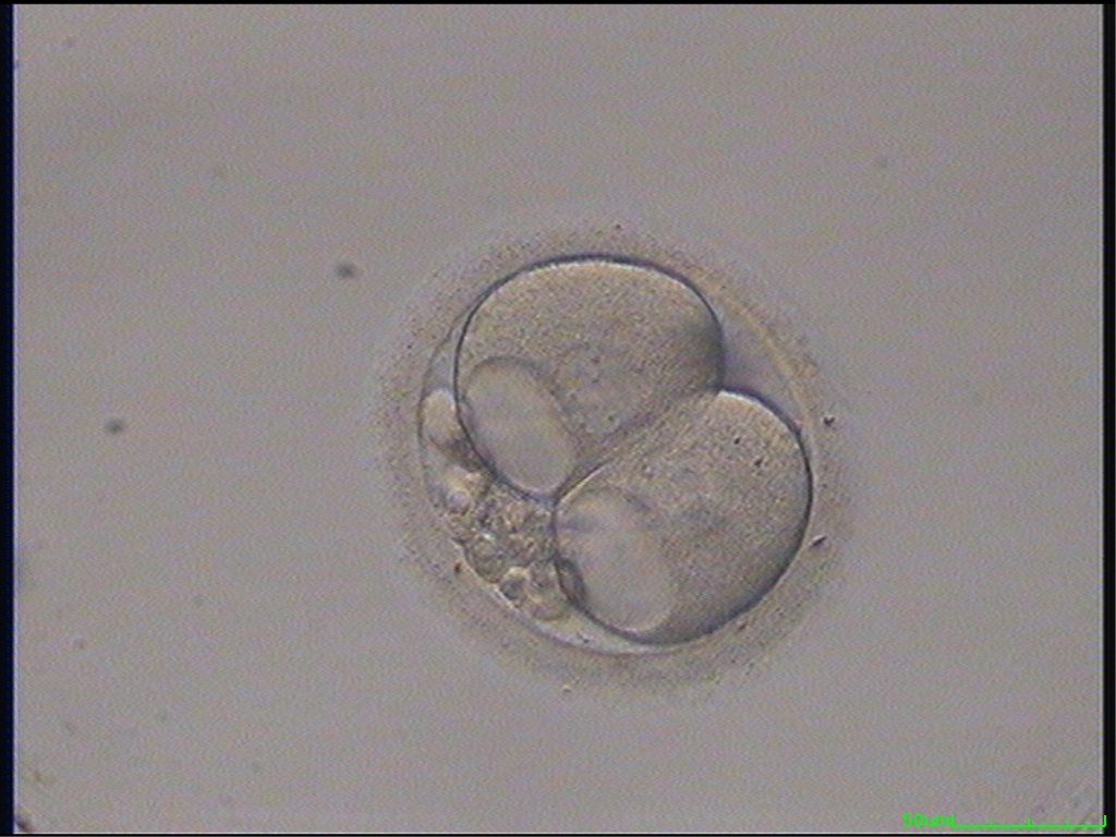

Figure 278
A 2-cell embryo with large vacuoles in both blastomeres and 15% concentrated fragmentation.

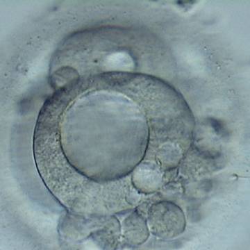
Figure 279
A 3-cell embryo with a large vacuole in the blastomere in the first plane in this view. A smaller vacuole is present in another blastomere. High fragmentation, about 40%, concentrated in one area.
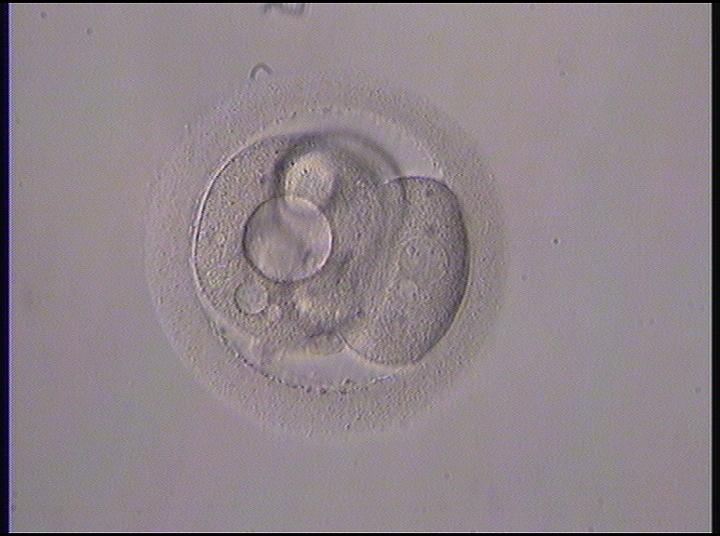
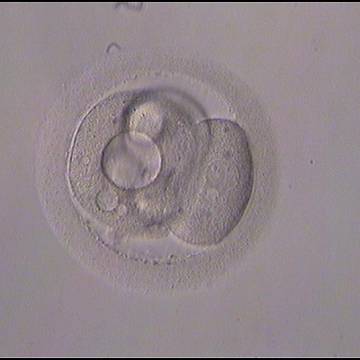
Figure 280
A 3-cell embryo with different sized blastomeress showing both large and small vacuoles.
Article references:
Alpha Scientists in Reproductive medicine and ESHRE Special Interest Group of Embryology. The Istanbul consensus workshop on embryo assessment: proceedings of an expert meeting. Hum Reprod 2011;26:1270-1283.
Abstract/FREE Full Text
Biggers JD, Racowksy C. The development of fertilized human ova to the blastocyst stage in KSOMAA medium: is a two-step protocol necessary? Reprod Biomed Online 2002;5:133-140.
Medline | Google Scholar
Hartshorne F. The embryo. Hum Reprod 2000;15:31-41.
FREE Full Text
Rienzi L, Ubaldi F, Minasi MG, Iacobelli M, Martinez F, Tesarik J, Greco E. Blastomere cytoplasmic granularity is unrelated to developmental potential of day 3 human embryos. J Assist Reprod Genet 2003;20:314-317.
CrossRef | Medline | Google Scholar
Ebner T, Moser M, Sommergruber M, Gaiswinkler U, Shebl O, Jesacher K, Tews G. Occurrence and developmental consequences of vacuoles throughout preimplantation development. Fertil Steril 2005a;83:1635-1640.
CrossRef | Medline | Web of Science | Google Scholar
Ebner T, Tews G, Sommergruber M, Moser M. Cytoplasmic pitting has a negative influence on implantation outcome. J Assist Reprod Genet 2005b;22:239-244.
CrossRef | Medline | Google Scholar
Van Blerkom J. Occurrence and developmental consequences of aberrant cellular organization in meiotically mature human oocytes after exogeneous ovariona hyperstimulation. J Electron Microsc Techn 1990;16:324-346.
CrossRef | Medline | Web of Science | Google Scholar
Veeck LL. An Atlas of Human Gametes and Conceptuses: An Illustrated Reference for Assisted Reproductive Technology. New York, USA: Parthenon Publishing; 1999. Preembryo grading and degree of cytoplasmic fragmentation; p. 46-51.
Google Scholar
