C. Oocyte size and shape
A critical oocyte size is necessary for resumption of meiosis (Otoi et al., 2000). At the beginning of oocyte growth, size is determined by strong adhesion between the oolemma and the inner zona surface (Tartia et al., 2009). Around ovulation GLYT1 is activated which mediates glycin accumulation which in turn acts as an osmolyte and thus controls cell volume (Baltz and Tartia, 2009).
The mean ovarian diameter of MII oocytes may vary substantially (Fig. 24) but it is not related to fertilization or developmental quality of human ICSI embryos at the cleavage stage of development (Romão et al., 2010). The situation is different with giant oocytes (Balakier et al., 2002; Rosenbusch et al., 2002). This type of oocyte has about twice the volume of a normal oocyte (about 200 µm) and is tetraploid before meiosis due to their origin, i.e. nuclear but no cytoplasmic division in an oogonium or cytoplasmic fusion of two oogonia. These mechanisms explain the binucleate appearance of prophase I giant eggs (Figs 25 and 26). These oocytes always contribute to digynic triploidy (Figs 27 and 28) and must never be transferred, although the presence of at least one giant oocyte in a cohort of retrieved eggs (Figs 29 and 30) has no effect on treatment outcome (Machtinger et al., 2011).
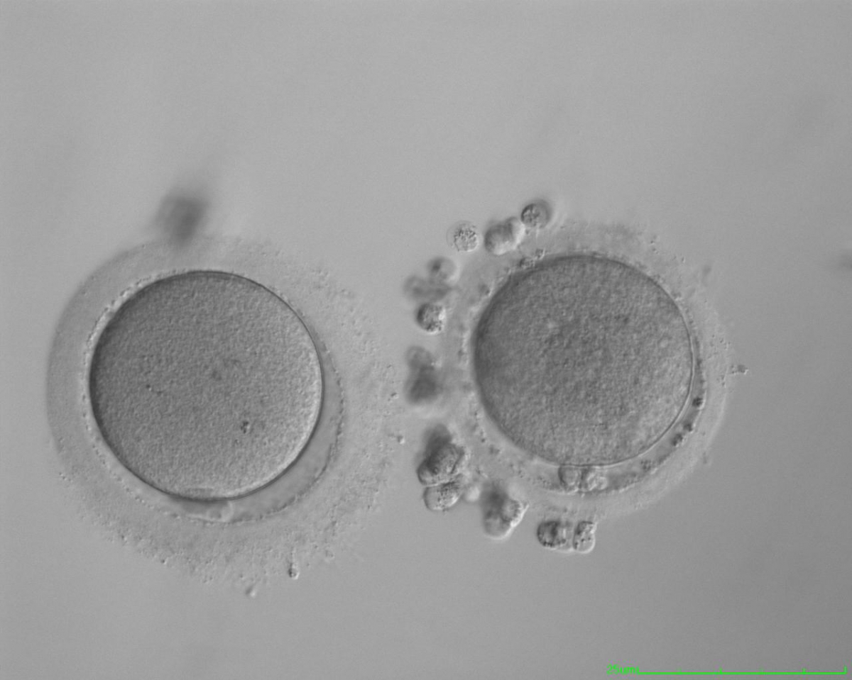

Figure 24
Small MII oocyte (right) next to a normal-sized MII oocyte (left) from the same cohort (200× magnification).
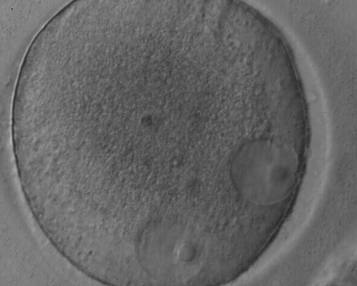

Figure 25
Giant oocyte with two apparent GVs (in an eccentric position). This is a tetraploid oocyte originating from the fusion of two separate oocytes (400× magnification).

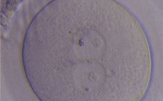
Figure 26
Giant oocyte with two apparent GVs (centrally located and juxtapposed). This tetraploid oocyte originates from the fusion of two separate oocytes and is usually tetraploid (400× magnification).
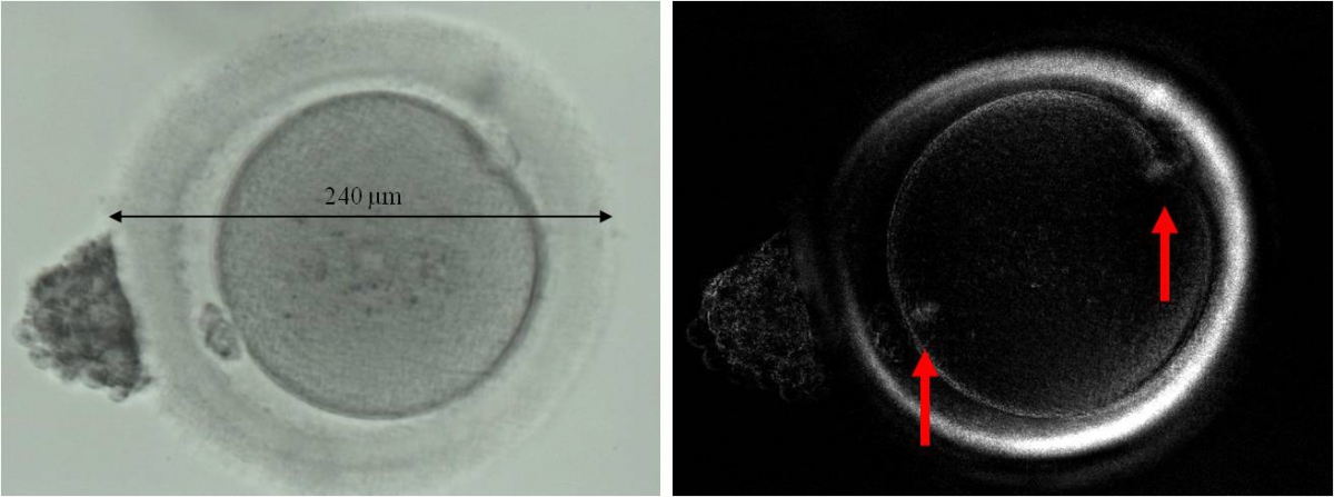

Figure 27
Giant MII oocyte visualized using bright field (left) and polarized light microscopy (right). The oocyte contains two distinct polar bodies and two distinct MSs at opposing poles of the oocyte.
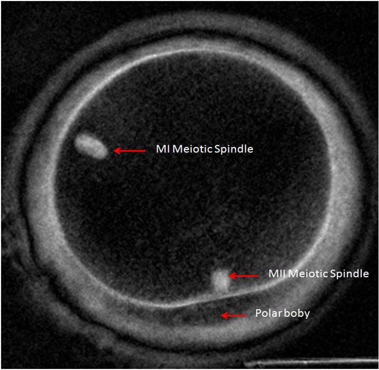
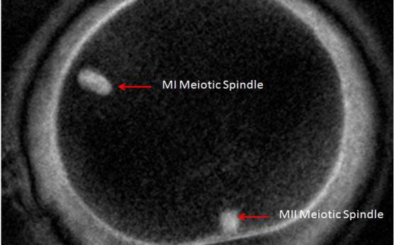
Figure 28
Giant MII oocyte visualized at high power using polarized light microscopy. The two distinct MSs can be observed in the cytoplasm. One MS is from the MI (10 o'clock position) and the other is from MII (6 o'clock position; 400× magnification).
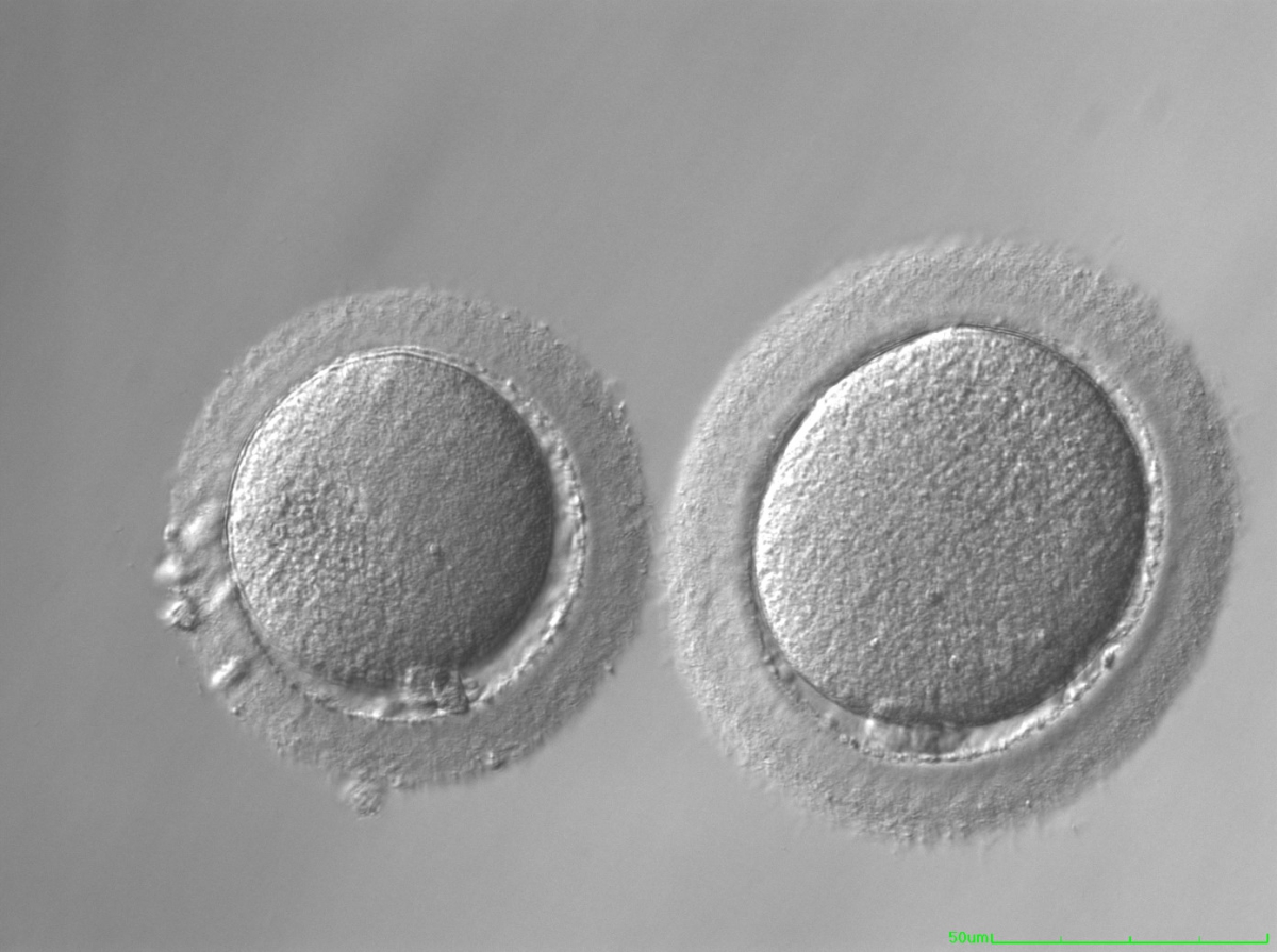
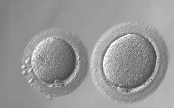
Figure 29
Giant oocyte (right) next to normal-sized oocyte (left; 200× magnification).
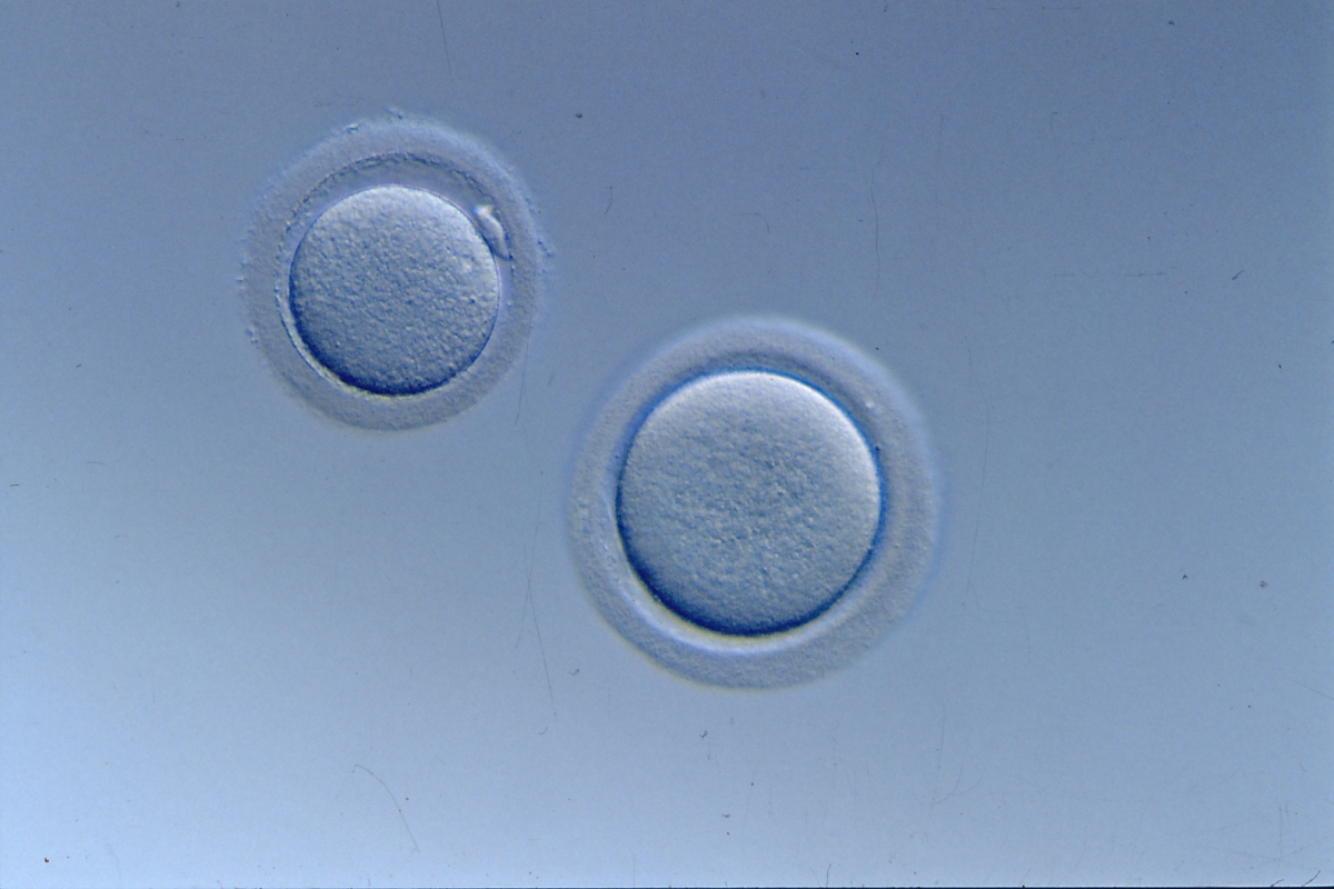
Figure 30
Giant oocyte (right) next to normal-sized oocyte (left; 200× magnification). No PB1 is visible in the giant oocyte.
It is evident that oocytes with extreme forms of shape anomaly exist (Figs 31 and 32; Paz et al., 2004; Esfandiari et al., 2005). Such ova have been shown to be fertilizable and may lead to the birth of healthy babies. While quantifying the degree of the elongation, some authors (Ebner et al., 2008) realized that the dimensions of the shape anomaly were neither correlated with fertilization nor embryo quality. However, when oocytes with ovoid zonae (Figs 33 and 34) develop, day 2 embryos show a flat array of blastomeres rather than the more traditional tetrahedral arrangement and further development is often delayed (Ebner et al., 2008).

Figure 31
Elongated MII oocyte inside an elongated ZP. Note the PVS appears relatively normal (400× magnification).

Figure 32
Elongated MII oocyte within a grossly distended and irregular ZP (200× magnification).
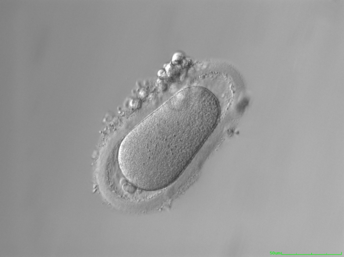

Figure 33
Ovoid MII oocyte. Note the ZP is also ovoid in appearance and the PVS is enlarged at both poles (200× magnification).
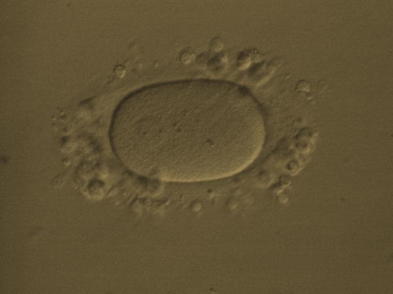

Figure 34
Ovoid MII oocyte. Note the ZP is also ovoid in appearance permitting the PVS to remain relatively normal (200× magnification).
Rarely, two oocytes can be found within the one follicular complex. Each oocyte is usually surrounded by a ZP but the ZP immediately between the two oocytes is commonly shared rather than duplicated (Fig. 35). It is not uncommon for these conjoined oocytes to show different nuclear maturational states. It has been suggested that such oocytes may play a role in producing dizygotic twins; however, even when both of the conjoined oocytes are mature it is rare that both fertilize and no pregnancies have been reported from such oocytes (Rosenbusch and Hancke, 2012).

Figure 35
Two oocytes enclosed within a single ZP (400× magnification).
Article references:
Balakier H, Bouman D, Sojecki A, Librach C, Squire JA. Morphological and cytogenetic analysis of human giant oocytes and giant embryos. Hum Reprod 2002;17:2394-2401.
Abstract/FREE Full Text
Baltz JM, Tartia AP. Cell volume regulation in oocytes and early embryos: connecting physiology to successful culture media. Hum Reprod Update 2009;16:166-176.
Medline | Web of Science | Google Scholar
Ebner T, Shebl O, Moser M, Sommergruber M, Tews G. Developmental fate of ovoid oocytes. Hum Reprod 2008;23:62-66.
Abstract/FREE Full Text
Esfandiari N, Ryan EAJ, Gotlieb L, Casper RF. Successful pregnancy following transfer of embryos from oocytes with abnormal zona pellucida and cytoplasm morphology. Reprod Biomed Online 2005;11:620-623.
CrossRef | Medline | Web of Science | Google Scholar
Machtinger R, Politch JA, Hornstein MD, Ginsburg ES, Racowsky C. A giant oocyte in a cohort of retrieved oocytes: does it have any effect on the in vitro fertilization cycle outcome? Fertil Steril 2011;95:573-576.
CrossRef | Medline | Google Scholar
Otoi T, Fujii M, Tanaka M, Ooka A, Suzuki T. Oocyte diameter in relation to meiotic competence and sperm penetration. Theriogenology 2000;54:534-542.
Google Scholar
Paz G, Amit A, Yavetz H. Case report: pregnancy outcome following ICSI of oocytes with abnormal cytoplasm and zona pellucida. Hum Reprod 2004;19:586-589.
Abstract/FREE Full Text
Romão GS, Araújo MC, de Melo AS, de Albuquerque Salles Navarro PA, Ferriani RA, Dos Reis RM. Oocyte diameter as a predictor of fertilization and embryo quality in assisted reproduction cycles. Fertil Steril 2010;93:621-625.
CrossRef | Medline | Google Scholar
Rosenbusch B, Hancke K. Conjoined human oocytes observed during assisted reproduction: description of three cases and review of the literature. Rom J Morphol Embryol 2012;53:189-192.
Medline | Google Scholar
Rosenbusch B, Schneider M, Gläser B, Brucker C. Cytogenetic analysis of giant oocytes and zygotes to assess their relevance for the development of digynic triploidy. Hum Reprod 2002;17:2388-2393.
Abstract/FREE Full Text
Tartia AP, Rudraraju N, Richards T, Hammer MA, Talbot P, Baltz JM. Cell volume regulation is initiated in mouse oocytes after ovulation. Development 2009;136:2247-2254.
Abstract/FREE Full Text
