A. Fertilization assessment
A.1 2PN
When assessments are performed 17 ± 1 h post-insemination, taking into consideration that pronuclear formation in IVF zygotes lags ∼1 h behind ICSI zygotes, normally fertilized oocytes should be spherical and have two polar bodies and two PNs (Figs 85–87). PNs should be juxtaposed, approximately the same size, centrally positioned in the cytoplasm with two distinctly clear, visible membranes (Fig. 88). The presence of an equal number and size of NPBs aligned at the PN junction has been correlated with increased embryo competence (Tesarik and Greco, 1999; Tesarik et al., 2000; Scott, 2003).
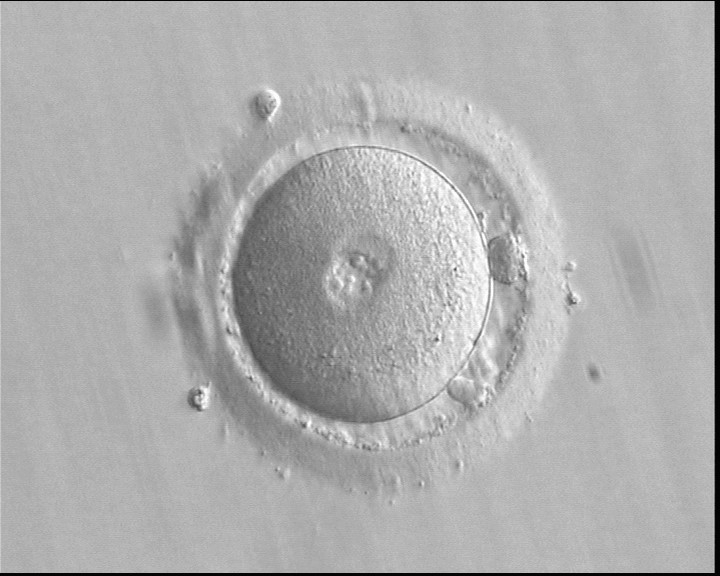
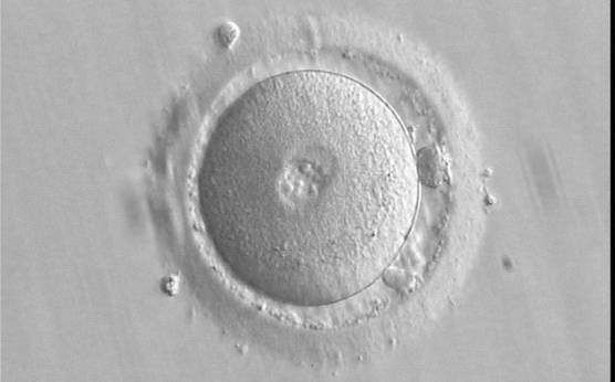
Figure 85
A zygote 16.5 h post-ICSI, having small-sized PNs with scattered NPBs and two visible polar bodies (400× magnification). The zona pellucida (ZP) appears regular; some debris is present in the perivitelline space (PVS). The cytoplasm is homogeneous and displays a clear cortical zone. It was transferred resulting in a clinical pregnancy followed by miscarriage.

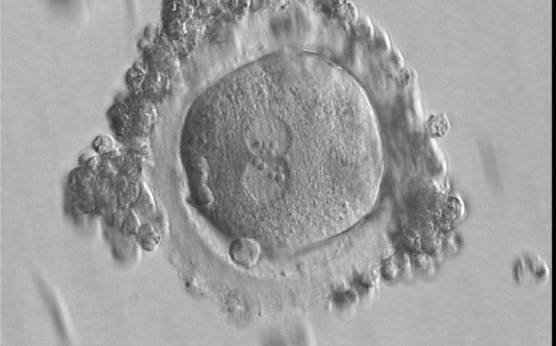
Figure 86
A slightly deformed zygote at 16.5 h after IVF with equal numbers of large-sized NPBs aligned at the PN junction (400× magnification). A great angle separates the two polar bodies. Some granulosa cells surround the ZP. It was transferred but failed to implant.
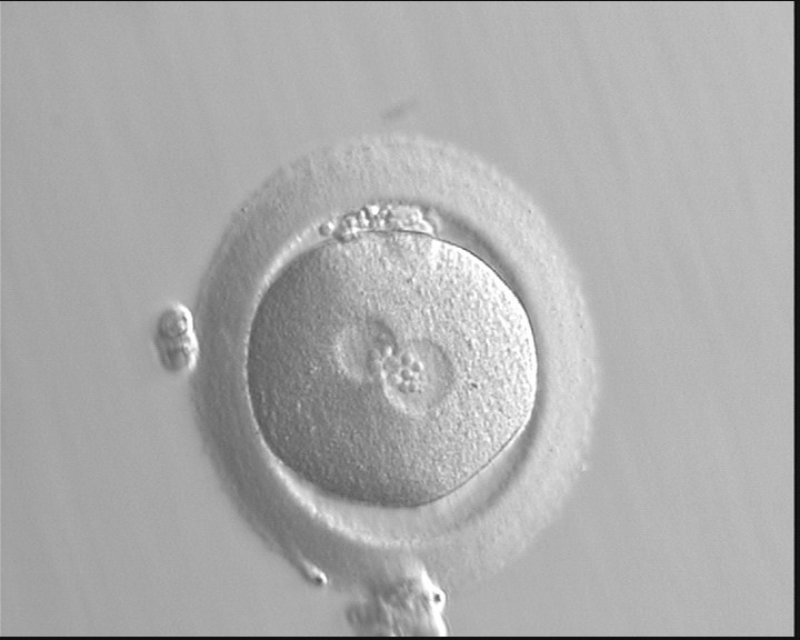
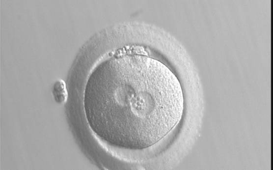
Figure 87
A zygote at 18.5 h generated by standard insemination using frozen/thawed ejaculated sperm (400× magnification). The two PNs are centrally located: one is slightly larger than the other. NPBs are of the same size, but different in number and are aggregated at adjacent borders of each PN. The ZP appears thick. It was transferred but failed to implant.

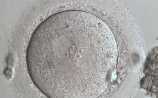
Figure 88
A zygote generated by ICSI with NPBs perfectly aligned at the junction of centrally located and juxtaposed PNs (600× magnification). Fragmented polar bodies are located in the longitudinal axis of the PNs. Category 1 (equivalent to Z1 score;Scott, 2003) was assigned following assessment. Debris appears to be present in the PVS. The cytoplasm is light-coloured with a clear cortical zone. It was transferred and implanted.
A.2 1PN
The incidence of one pronucleates is around 1% after IVF or ICSI and may be parthenogenetic in origin as suggested by chromosomal analysis that shows a haploid set in approximately half of the studied oocytes (Plachot, 2000). In some cases, only one polar body is extruded into the perivitelline space (PVS) (Fig. 89).
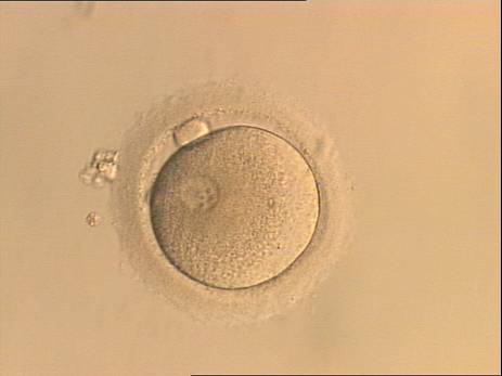

Figure 89
A single pronucleate oocyte displaying only one PN and a single polar body observed 16 h post-ICSI (200× magnification). The observation was repeated 17.5, 20 and 22 h post-ICSI and did not show significant variation in the PN size or position.
The presence of 1PN can also be a result of errors in the fertilization process with asynchrony in PN formation or PN fusion. In these cases, the resulting oocyte could have a diploid set of chromosomes, and two polar bodies are normally observed (Figs 90–93). The transfer of these oocytes could be considered, but the incidence of aneuploidy in the resulting embryos has been reported to be significantly higher compared with embryos derived from 2PN oocytes (Yan et al., 2010).


Figure 90
A zygote observed 15 h post-IVF displaying a single, large-sized PN and two polar bodies (400× magnification).
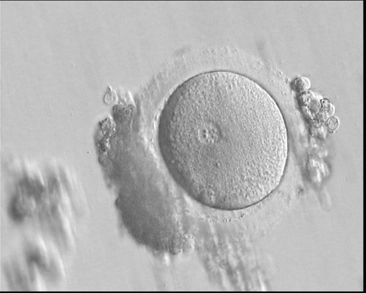

Figure 91
A zygote 17 h post-IVF showing a single PN with NPBs of different size and two polar bodies (400× magnification).
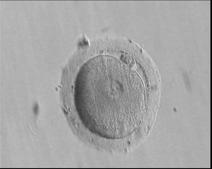

Figure 92
A single, large PN and two polar bodies (partially overlapping) are present in this oocyte observed 16 h and 45 min post-IVF (400× magnification). Four large-sized NPBs are visible. The resulting embryo was transferred, but failed to implant.


Figure 93
A zygote generated by ICSI showing a single PN and two polar bodies separated by some distance (600× magnification).
A.3 ≥3PN
The formation of triploid zygotes differs in origin, depending on whether they are generated by ICSI or IVF. Only 1% of oocytes after ICSI result in tripronuclear zygotes and are digynic due to failure in extrusion of the second polar body (Fig. 94). An exception is represented by giant oocytes (Fig. 95) that follow different patterns of extrusion due to their generally diploid condition (see Chapter One).
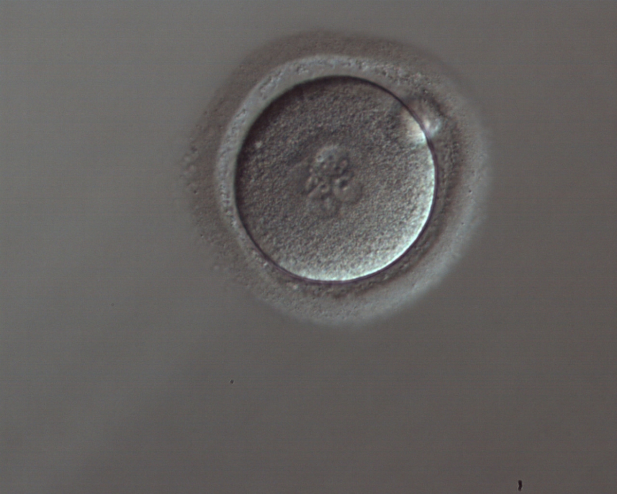
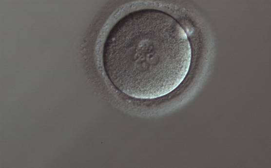
Figure 94
A zygote generated by ICSI displaying four PNs of approximately the same size and two of smaller size (150× magnification). Only one polar body is visible.
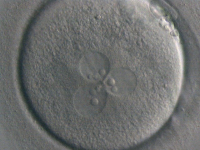
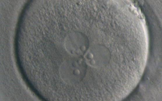
Figure 95
A zygote displaying 3PNs with large-sized NPBs (400× magnification). One of the three PNs is slightly bigger than the others. The zygote was generated by ICSI performed on a giant oocyte.
Diandry is the most probable cause of tripronucleates following IVF and occurs in ∼5% of inseminated oocytes (Fig. 96). This condition arises from the entry of two spermatozoa into the cytoplasm owing to the incapacity of the oocyte to trigger protection against polyspermy. The second polar body is normally extruded and cleavage often occurs. In some cases, the presence of multiple PNs could be due to failed cytokinesis (Figs 94 and 97) or to penetration by a binucleate spermatozoon (Fig. 98).


Figure 96
A zygote displaying 3PNs of approximately the same size with large-sized NPBs, partly overlapped and aligned in the middle of the oocyte (400× magnification). It was generated by IVF and shows two polar bodies.
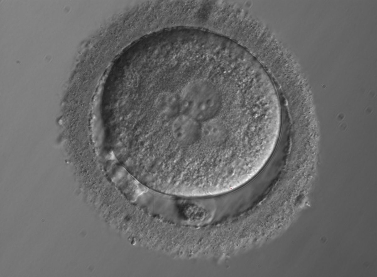
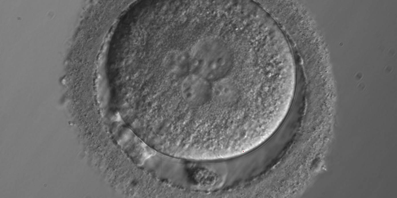
Figure 97
A zygote with 5PNs, halo cytoplasm, fragmented polar bodies, oval shape and dark ZP (400× magnification). It was warmed after vitrification.
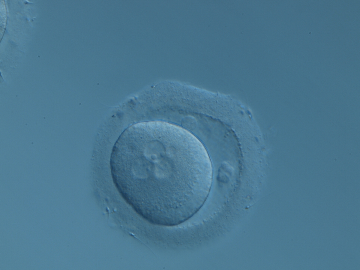

Figure 98
A zygote displaying 3PNs after IVF with a small fragment adjacent to the PNs (200× magnification). There are two polar bodies in a large PVS and a thick ZP.
Article references:
Plachot M. Fertilization. Hum Reprod 2000;15(Suppl. 4):19-30.
FREE Full Text
Scott L. Pronuclear scoring as a predictor of embryo development. Reprod Biomed Online 2003;6:201-214.
CrossRef | Medline | Google Scholar
Tesarik J, Greco E. The probability of abnormal preimplantation development can be predicted by a single static observation on pronuclear stage morphology. Hum Reprod 1999;14:1318-1323.
Abstract/FREE Full Text
Tesarik J, Junca AM, Hazout A, Aubriot FX, Nathan C, Cohen-Bacrie P, Dumont-Hassan M. Embryos and high implantation potential after intracytoplasmic sperm injection can be recognized by a simple, non- invasive examination of pronuclear morphology. Hum Reprod 2000;15:1396-1399.
Abstract/FREE Full Text
Yan J, Li Y, Shi Y, Feng HL, Gao S, Chenz Z. Assessment of sex chromosomes of human embryos arising from monopronucleus zygotes in in vitro fertilization and intracytoplasmic sperm injection cycles of Chinese women. Gynecol Obstet Invest 2010;69:20-23.
CrossRef | Medline | Google Scholar
