A. Cell numbers
The developmental stage of an embryo, defined as the number of blastomeres on Days 1, 2 or 3 after insemination is an essential predictive factor for subsequent implantation and pregnancy rates (1 cell to >10 cells; Figs 211–222). For assessment of embryo cleavage (numbers of blastomeres), the currently accepted observation schedule for optimal cleavage rates was defined at the Istanbul consensus workshop to be: Day 1 (26 ± 1 h post-ICSI, 28 ± 1 h post-IVF), 2-cells; Day 2 (44 ± 1 h), 4-cells and Day 3 (68 ± 1 h), 8-cells (Alpha Scientists in Reproductive Medicine and ESHRE Special Interest Group of Embryology, 2011). Early cleavage (Figs 211 and 212), i.e. the first mitosis occurring before 26±1 h (ICSI) and 28±1 h (IVF) respectively, has been shown to correlate with numbers of good quality embryos, blastocyst development and pregnancy rates (Lundin et al., 2001; Fenwick et al., 2002). A number of studies have shown that the transfer of 4-cell embryos on Day 2 of culture (Fig. 215) results in significantly higher implantation and pregnancy rates compared with the transfer of embryos with either lower (Figs 213 and 214) or higher (Fig. 216) cell numbers (Thurin et al., 2005; Holte et al., 2007; Scott et al., 2007).
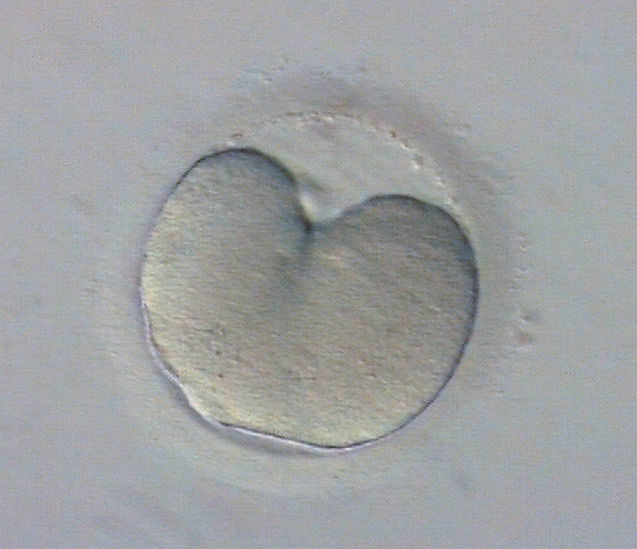

Figure 211
A zygote undergoing first mitosis which has not been completed by 25 h post-insemination. The mitotic groove can be seen. This embryo should still be assessed as a 1-cell embryo.
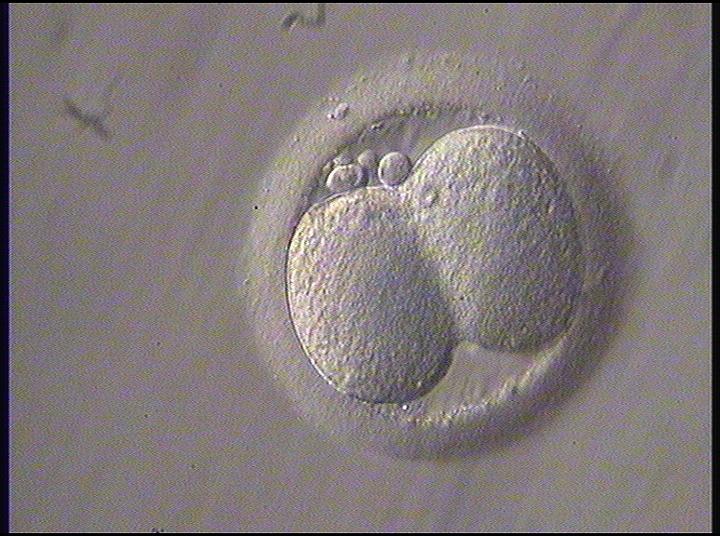

Figure 212
The first embryo division nearing completion but the two daughter cells are still not separated. This should be assessed as a 2-cell embryo.
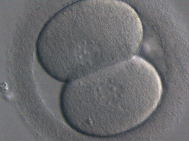

Figure 213
A 2-cell embryo with evenly sized blastomeres each containing one nucleus. Generated by ICSI but was not transferred.
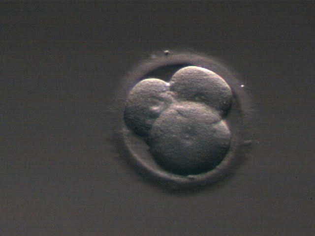

Figure 214
A 3-cell embryo with one larger blastomere and two smaller blastomeres, i.e. it has a stage-specific cell size (see Section C). The embryo was generated by ICSI but was not transferred.
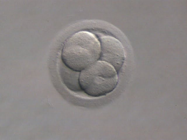

Figure 215
A 4-cell embryo with four evenly sized blastomeres each one containing one nucleus. Generated by ICSI. The embryo was transferred and implanted.
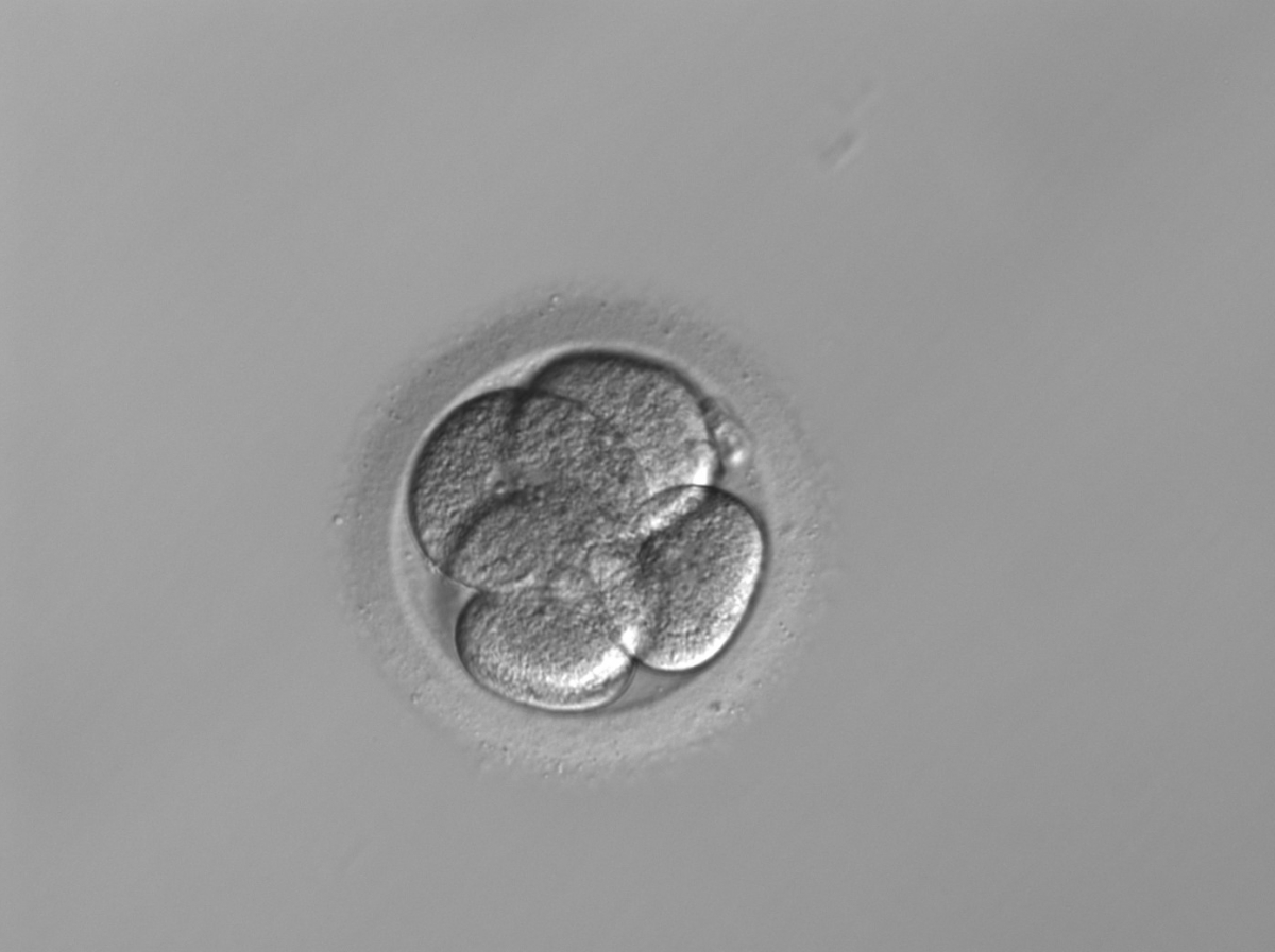

Figure 216
A 5-cell embryo where one cell is slightly out of focus. There is a slight difference in cell size but not significant enough to be called an uneven sized embryo. It was generated by ICSI and cryopreserved.
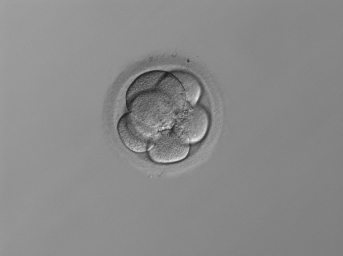

Figure 217
A 6-cell embryo with four small and two larger blastomeres, i.e. it has a stage-specific cell size (see Section C). The embryo was generated by IVF and cryopreserved.
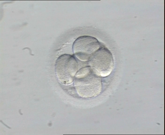

Figure 218
A 7-cell embryo in which four of the blastomeres show a single nucleus. The embryo was generated by ICSI, it was transferred and implanted.
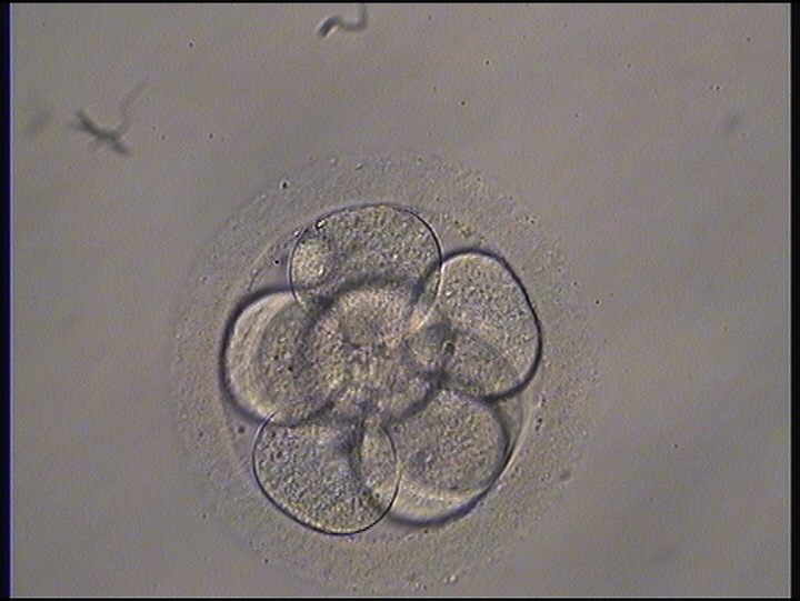

Figure 219
An 8-cell embryo with evenly sized blastomeres with no visible nuclei. It was generated by ICSI and transferred but the outcome is unknown.
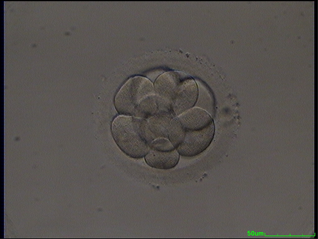

Figure 220
A cryopreserved 9-cell embryo, warmed on Day 3. One blastomere is slightly larger and one blastomere is slightly smaller than the others. It was generated by ICSI and transferred but failed to implant.
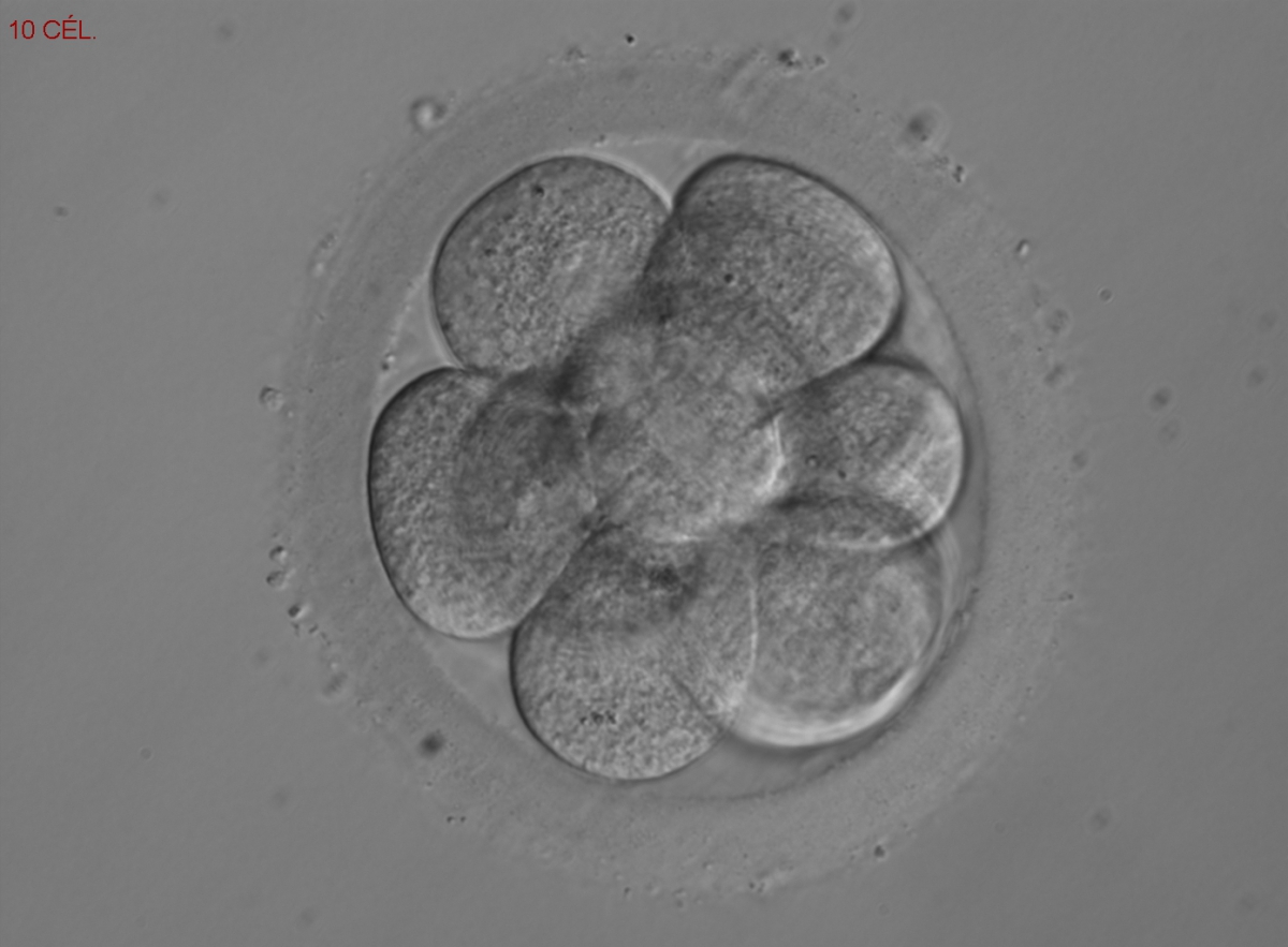

Figure 221
A 10-cell embryo with visible nuclei in some blastomeres. It was generated by ICSI and cryopreserved.
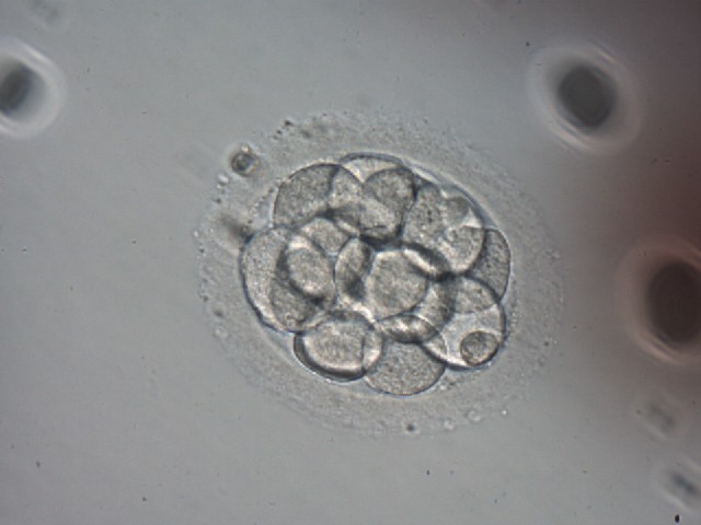

Figure 222
An embryo with more than 10 cells on Day 4. This embryo has not compacted which is unusual at this late stage. Generated by ICSI and cryopreserved.
Correspondingly, several studies have shown that for Day 3 transfers, implantation and live birth rates are positively correlated with an increase in cell number on Day 3, with the 8-cell stage (having been a 4-cell embryo on Day 2) having the highest rates (van Royen et al., 1999; Racowsky et al., 2011). The cleavage stage of the embryo at the time of transfer also seems to have a role in predicting early pregnancy loss. Hourvitz et al. (2006) found that five or less blastomeres in the best embryo transferred on Day 3 was correlated with early pregnancy loss. A correlation between cell numbers at distinct observation time points and chromosomal errors has also been reported. It was shown by Munné (2006) that Day 2 embryos with 4 cells had the lowest rate of chromosomal errors, while Magli et al. (2007) showed the same to be true for embryos with 7- to 8 cells on Day 3 (Figs 218 and 219). The same pattern was observed by Finn et al. (2010) who described a higher rate of euploidy in embryos with seven to eight blastomeres on Day 3 compared with both six (Fig. 217) or less than six blastomeres and nine (Fig. 220) or more than nine (Figs 221 and 222) blastomeres.
Article references:
Alpha Scientists in Reproductive medicine and ESHRE Special Interest Group of Embryology. The Istanbul consensus workshop on embryo assessment: proceedings of an expert meeting. Hum Reprod 2011;26:1270-1283.
Abstract/FREE Full Text
Fenwick J, Platteau P, Murdoch AP, Herbert M. Time from insemination to first cleavage predicts developmental competence of human preimplantation embryos in vitro. Hum Reprod 2002;17:407-412.
Abstract/FREE Full Text
Finn A, Scott L, O'leary T, Davies D, Hill J. Sequential embryo scoring as a predictor of aneuploidy in poor-prognosis patients. Reprod Biomed Online 2010;21:381-390.
CrossRef | Medline | Web of Science | Google Scholar
Holte J, Berglund L, Milton K, Garello C, Gennarelli G, Revelli A, Bergh T. Construction of an evidence-based integrated morphology cleavage embryo score for implantation potential of embryos scored and transferred on day 2 after oocyte retrieval. Hum Reprod 2007;22:548-557.
Abstract/FREE Full Text
Hourvitz A, Lerner-Geva L, Elizur SE, Baum M, Levron J, David B, Meirow D, Yaron R, Dor J. Role of embryo quality in predicting early pregnancy loss following assisted reproductive technology. Reprod Biomed Online 2006;13:504-509.
CrossRef |Medline | Web of Science | Google Scholar
Lundin K, Bergh C, Hardarson T. Early embryo cleavage is a strong indicator of embryo quality in human IVF. Hum Reprod 2001;16:2652-2657.
Abstract/FREE Full Text
Magli MC, Gianaroli L, Ferraretti AP, Lappi M, Ruberti A, Farfalli V. Embryo morphology and development are dependent on the chromosomal complement. Fertil Steril 2007;87:534-540.
CrossRef | Medline | Web of Science | Google Scholar
Munné S. Chromosome abnormalities and their relationship to morphology and development of human embryos. Reprod Biomed Online 2006;12:234-253.
Medline | Web of Science | Google Scholar
Racowsky C, Stern JE, Gibbons WE, Behr B, Pomeroy KO, Biggers JD. National collection of embryo morphology data into Society for Assisted Reproductive Technology Clinic Outcomes Reporting System: associations among day 3 cell number, fragmentation and blastomere asymmetry, and live birth rate. Fertil Steril 2011;95:1985-1989.
CrossRef | Medline | Google Scholar
Scott L, Finn A, O'Leary T, McLellan S, Hill J. Morphologic parameters of early cleavage-stage embryos that correlate with fetal development and delivery: prospective and applied data for increased pregnancy rates. Hum Reprod 2007;22:230-240.
Abstract/FREE Full Text
Thurin A, Hardarson T, Hausken J, Jablonowska B, Lundin K, Pinborg A, Bergh C. Predictors of ongoing implantation in IVF in a good prognosis group of patients. Hum Reprod 2005;20:1876-1880.
Abstract/FREE Full Text
Van Royen E, Mangelschots K, De Neubourg D, Valkenburg M, Van de Meerssche M, Ryckaert G, Eestermans W, Gerris J. Characterization of a top quality embryo, a step towards single-embryo transfer. Hum Reprod 1999;14:2345-2349.
Abstract/FREE Full Text
