B. Pronuclear size
B.1 Normal
Some studies have demonstrated that the size of PNs depends on gamete factors. Decondensation of the tightly compacted sperm chromatin is a crucial step in fertilization that includes the replacement of protamines with histones under the control of oocyte-decondensing factors. Therefore, the male PN size depends both on the sperm nuclear structure and on the oocyte's capacity of inducing decondensation by releasing appropriate levels of glutathione.
PNs normally appear to be similar in size, although the female PN that is often located towards the second polar body can be slightly smaller (Figs 99 and 100). However, PN formation and rotation is a dynamic process as demonstrated by time-lapse video recording, so positioning and morphology of PNs are strictly time dependent (Fig. 101).
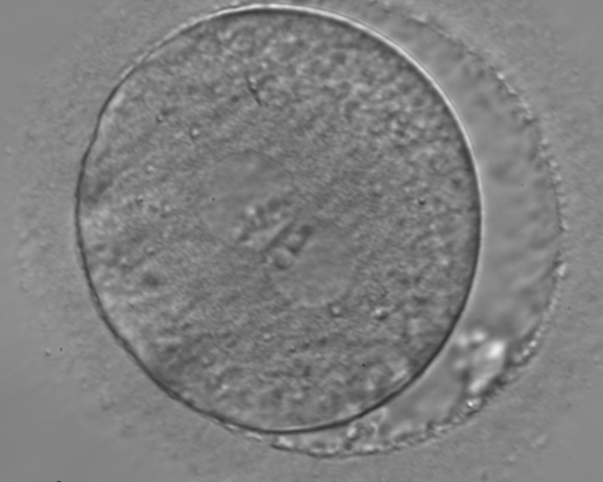
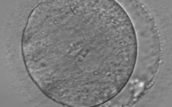
Figure 99
A zygote observed 18 h post-ICSI (400× magnification). The 2PNs are centrally located and juxtaposed in the cytoplasm (peripherally granular), of approximately the same size, and exhibit inequality in the number and size of NPBs. The PN on the right demonstrates fewer but larger nucleoli. Some debris appears to be present in the slightly increased PVS. Transferred on Day 3 (eight cells) along with two other embryos to a patient who delivered a healthy baby boy.

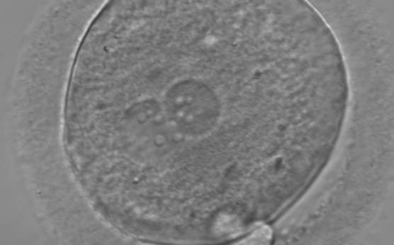
Figure 100
A zygote with equal numbers of large-sized NPBs scattered with respect to the PN junction (400× magnification). PNs are juxtaposed and slightly eccentric. The two polar bodies are located in a plane that is parallel to the longitudinal axis of the PNs.

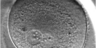
Figure 101(a)
A zygote with changes in PN pattern, particularly with respect to the position in the cytoplasm and the NPBs' location over time at (a) 11.7h after ICSI (400× magnification).
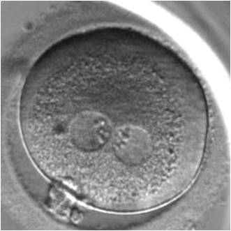
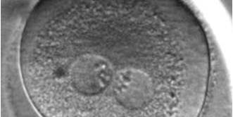
Figure 101(b)
15.4 h after ICSI (400× magnification).
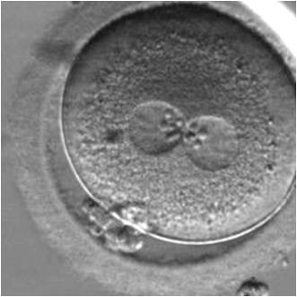
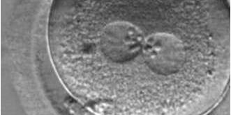
Figure 101(c)
18.3 h after ICSI (400× magnification).

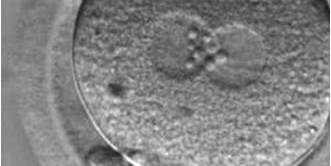
Figure 101(d)
28.3h after ICSI (400× magnification).
B.2 Small
The size of PNs depends on the time of observation with an increase in size of close to 50% from abuttal to 17 h post-ICSI (Payne et al., 1997). The presence of PNs with a diameter smaller than normal (Figs 102–104) at the normal time of fertilization check could be an indicator of delayed fertilization possibly due to oocyte immaturity, or defects in the gametes. Nevertheless, implantation can occur as a result of transfer of these zygotes (Fig. 103).
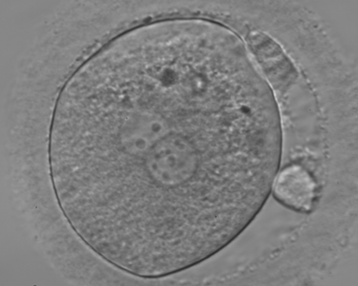
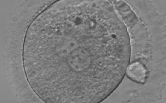
Figure 102
A zygote generated by ICSI and observed 18 h post-insemination (400× magnification). PNs are slightly smaller than normal and exhibit inequality in the number and the distribution of NPBs. Polar bodies are larger than the normal size.
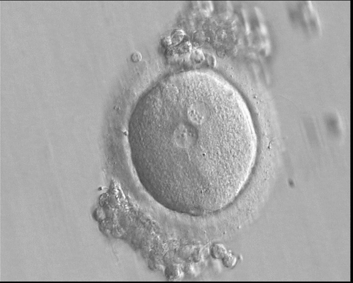
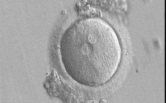
Figure 103
A zygote generated by IVF using frozen/thawed ejaculated sperm and observed 16 h post-insemination (400× magnification). PNs are smaller than normal and are not exactly positioned in the centre of the oocyte. Small-sized NPBs are aligned at the PN junction. It was transferred and implanted.
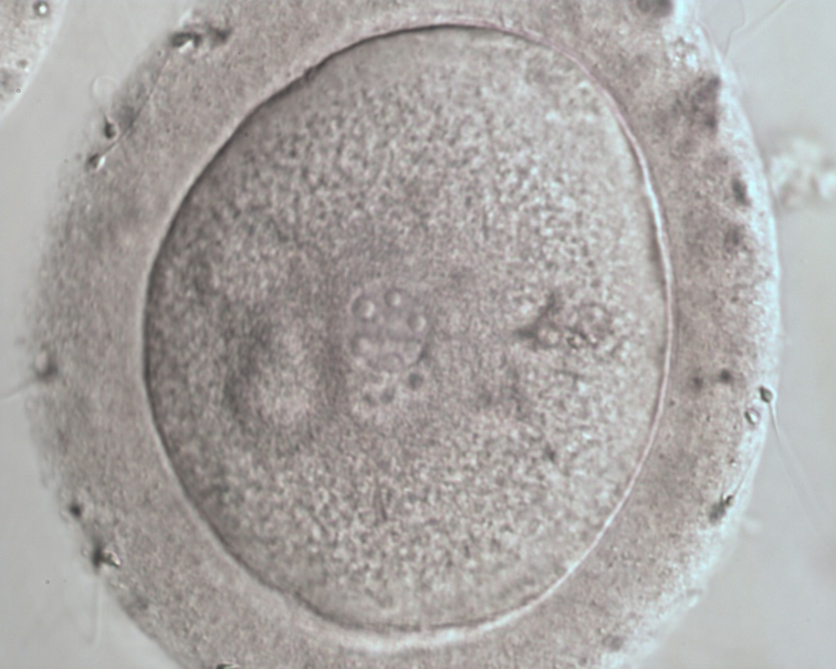
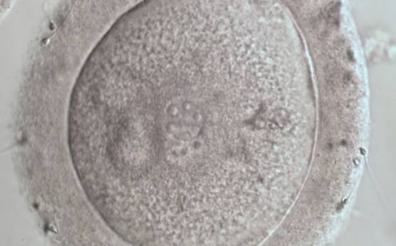
Figure 104
A zygote displaying two small PNs partly overlapping in this view (600× magnification). NPBs are large in size, equal in numbers and scattered in the two PNs. The ZP appears thickened and the PVS almost absent.
B.3 Differential in size
Significant differences (>4 μm) between PN size (Figs 105–108), or the presence of micronuclei or fragmented PNs (Figs 109 and 110) are considered to be abnormal and are associated with chromosomal abnormalities and major loss of developmental potential (Munné and Cohen, 1998; Scott et al., 2000; Nagy et al., 2003; Scott et al., 2007; Alpha Scientists in Reproductive medicine and ESHRE Special Interest Group of Embryology, 2011). Clinical studies have shown a high correlation between zygotes observed at 16–18 h post-insemination displaying large differences in PN size and their capability to maintain viability and development both in vivo and in vitro (Sadowy et al., 1998; Scott et al., 2000). Therefore, when performing embryo selection these embryos should be avoided for transfer.

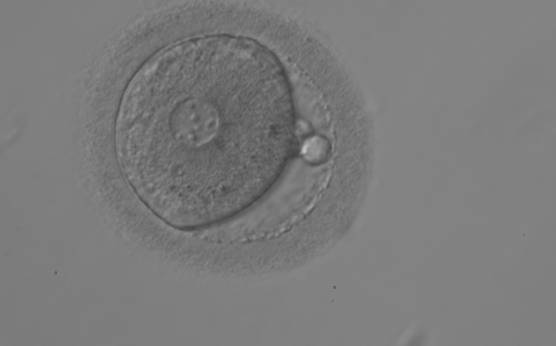
Figure 105
A zygote observed 18 h post-ICSI displaying very unequal-sized juxtaposed PNs, with the smaller PN being less visible (200× magnification). Two polar bodies and small-sized NPBs are scattered in both PNs.

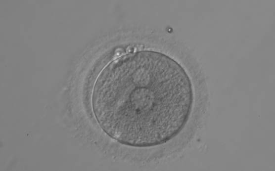
Figure 106
A zygote displaying two polar bodies and unequal-sized juxtaposed PNs and inequality in number and alignment of small-sized NPBs (200× magnification).
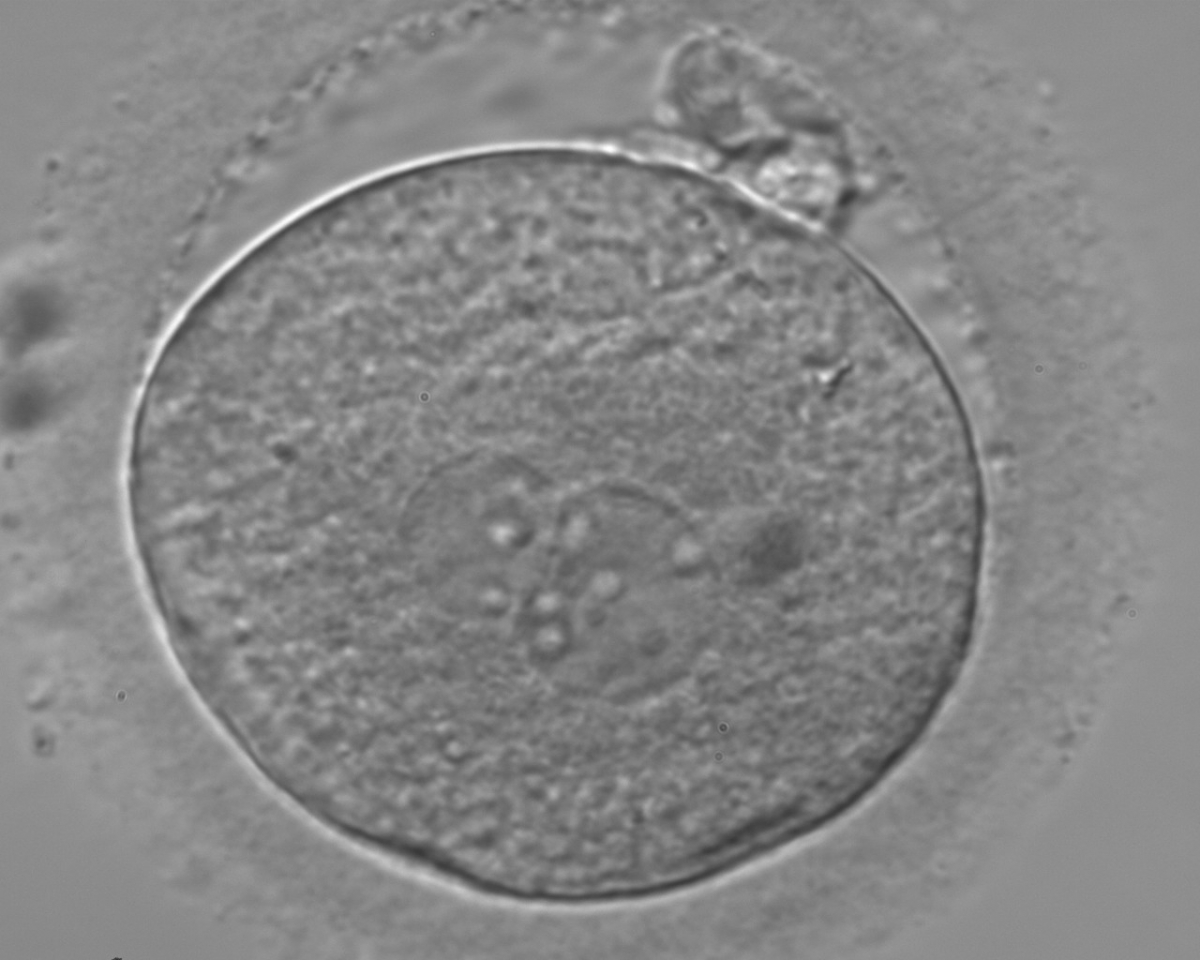
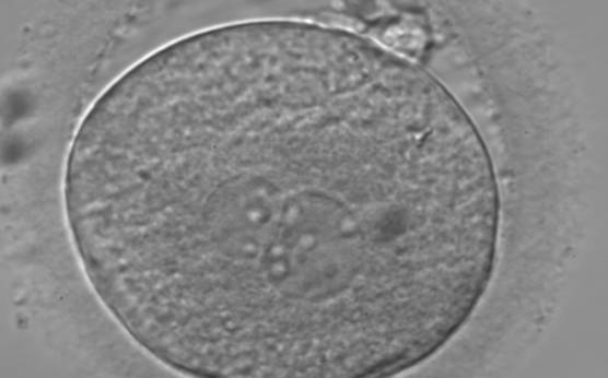
Figure 107
A zygote observed 18 h post-insemination displaying very unequal-sized PNs and inequality in number and alignment of NPBs (400× magnification). It was transferred on Day 3 (seven cells) along with two other embryos. The implantation result is therefore unknown. However, the patient delivered a healthy baby boy.
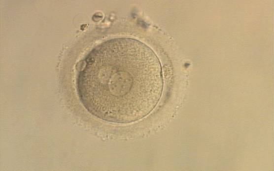
Figure 108
A zygote generated by ICSI with one large and one normal-sized PN (200× magnification). The two polar bodies are at opposite sides of the oocyte.
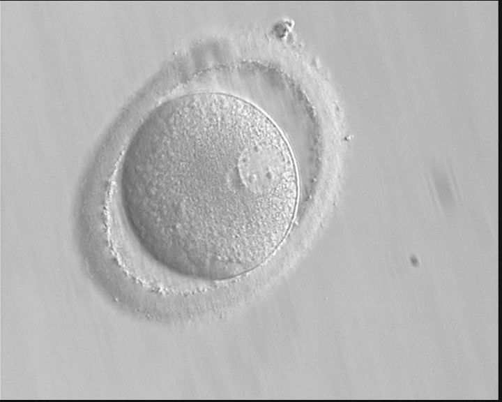
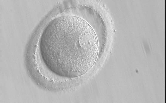
Figure 109
A zygote after PB biopsy showing peripheral PNs that are very different in size with one larger and one smaller than the normal size (400× magnification). The ZP is oval in shape.
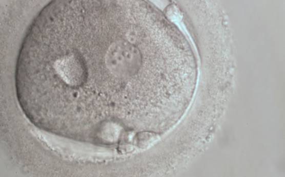
Figure 110
A zygote 18 h post-ICSI displaying very unequal-sized, juxtaposed PNs and inequality in number and alignment of NPBs (600× magnification). A vacuole-like structure is present in the cytoplasm. Fragments are visible in the PVS, not easily discernible from the polar bodies in this view.
Article references:
Alpha Scientists in Reproductive Medicine and ESHRE Special Interest Group of Embryology. The Istanbul consensus workshop on embryo assessment: proceedings of an expert meeting. Hum Reprod 2011;26:1270-1283.
Abstract/FREE Full Text
Munné S, Cohen J. Chromosome abnormalities in human embryos. Hum Reprod Update 1998;4:842-855.
Abstract/FREE Full Text
Nagy ZP, Dozortsev D, Diamond M, Rienzi L, Ubaldi F, Abdelmassih R, Greco E. Pronuclear morphology evaluation with subsequent evaluation of embryo morphology significantly increases implantation rates. Fertil Steril 2003;80:67-74.
Medline | Web of Science | Google Scholar
Payne D, Flaherty SP, Barry MF, Matthews CD. Preliminary observations on polar body extrusion and pronuclear formation in human oocytes using time-lapse video cinematography. Hum Reprod 1997;12:532-541.
Abstract/FREE Full Text
Sadowy S, Tomkin G, Munné S, Ferrara-Congedo T, Cohen J. Impaired development of zygotes with uneven pronuclear size. Zygote 1998;6:137-141.
CrossRef | Medline | Web of Science | Google Scholar
Scott L, Alvero R, Leondires M, Miller B. The morphology of human pronuclear embryos is positively related to blastocyst development and implantation. Hum Reprod 2000;15:2394-2403.
Abstract/FREE Full Text
Scott L, Finn A, O'Leary T, McLellan S, Hill J. Morphologic parameters of early cleavage-stage embryos that correlate with fetal development and delivery: prospective and applied data for increased pregnancy rates. Hum Reprod 2007;22:230-240.
Abstract/FREE Full Text
