A. Cumulus-Enclosed Oocytes
During follicular antrum formation, granulosa cells (GCs) differentiate into mural GCs, lining the follicular wall, and CCs, surrounding the oocyte. Within the cumulus mass, CCs in close contact with the oocyte (corona cells) develop cytoplasmic projections which cross the ZP and form gap junctions with the oolemma. This organized structure is called the cumulus–oocyte complex (COC; Fig. 1; Albertini et al., 2001). In natural spontaneous cycles, oocyte nuclear maturation runs parallel to the gradual FSH-dependent expansion of the cumulus and corona cells, whereas this synchrony may be disturbed in stimulated cycles (Laufer et al., 1984). Immature COCs (Fig. 2), commonly retrieved from small follicles during in vitro maturation (IVM) cycles, show a typically unexpanded cumulus with multilayers of compact GCs adhering to the ZP of an immature oocyte at prophase I [germinal-vesicle stage (GV); Figs 3 and 4]. IVM of such immature COCs aims for expansion of CCs and oocyte nuclear maturation.
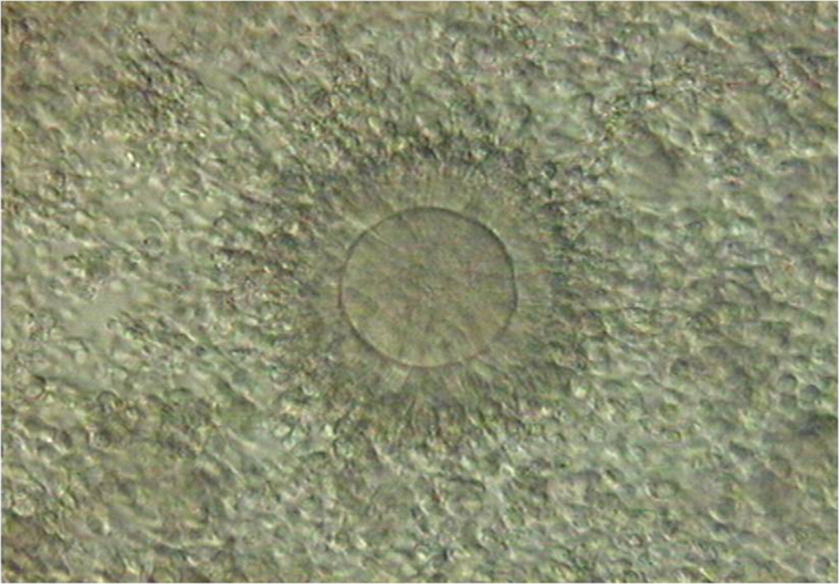
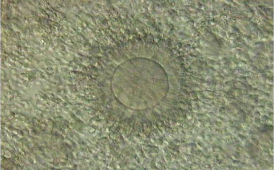
Figure 1. Cumulus–oocyte complex obtained following ovarian stimulation. The oocyte is typically surrounded by an expanded cumulus corona cell complex. Note the outer CCs separated from each other by extracellular matrix and the corona cells immediately adjacent to the oocyte becoming less compact and radiating away from the ZP. PB1 is located at the 1 o'clock position.
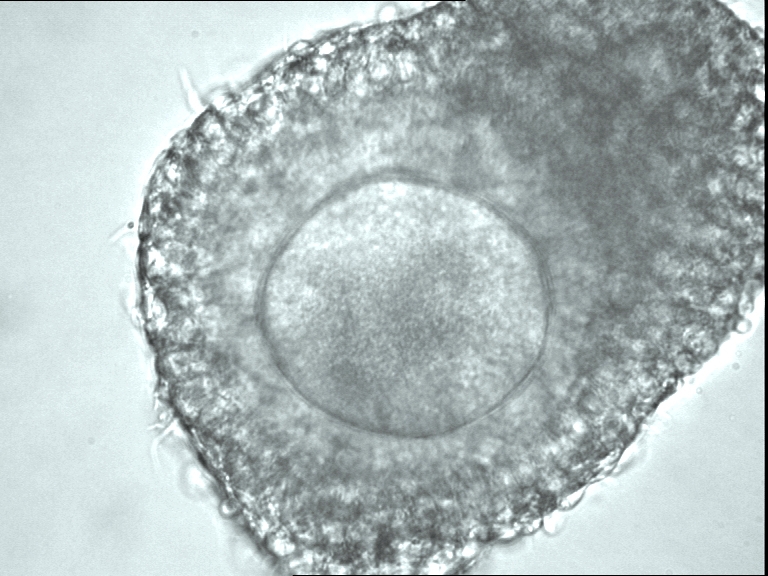
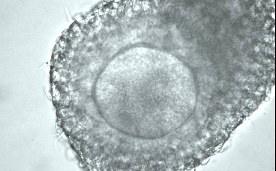
Figure 2
A cumulus–oocyte complex recovered from an IVM cycle showing an oocyte surrounded by unexpanded, compact cumulus and corona cells.
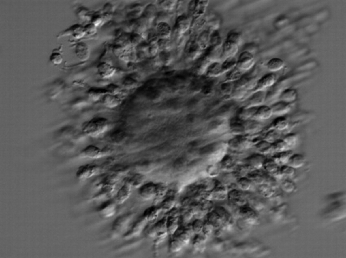
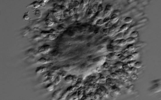
Figure 3. A cumulus–oocyte complex recovered from an IVM cycle. The immature GV oocyte is surrounded by compact GCs. The nucleolus in the GV is visible at the 10 o'clock position.
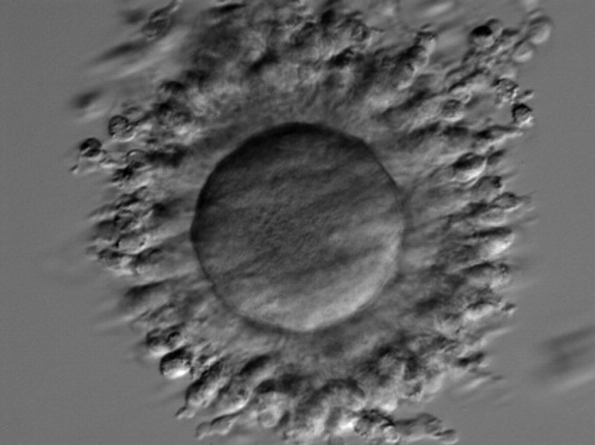
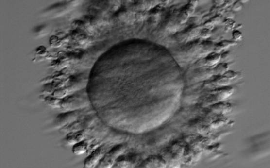
Figure 4. A cumulus–oocyte complex recovered from an IVM cycle. The immature oocyte has a GV at the 3 o'clock position with the nucleolus towards the centre of the oocyte. Compact GCs surround the oocyte.
In stimulated cycles, 34–38 h after triggering ovulation, a typical mature pre-ovulatory COC displays radiating corona cells surrounded by the expanded, loose mass of CCs (Fig. 5). In the majority of expanded COCs, oocytes are mature at the MII stage, although it is possible after gonadotrophin stimulation to find in a mucified cumulus and radiating corona cells an immature oocyte at the GV or metaphase I (MI) stage (Fig. 6). It is common, in stimulated cycles, to recover COCs with an expanded cumulus cell mass but compact, non-radiating corona cells (Figs 7a, 8a and 9a). Indeed, at recovery, the presence of the surrounding cumulus and corona cells usually prevents identification of the PBI in the PVS, an indicator of successful completion of meiosis I with arrest at the MII stage of development (Fig. 1). In preparation for ICSI, oocyte denudation is performed via enzymatic action of hyaluronidase (Figs 7b , 8b and 9b) and mechanical pipetting, allowing the accurate determination of the oocyte nuclear status (Figs 7c , 8c and 9c).
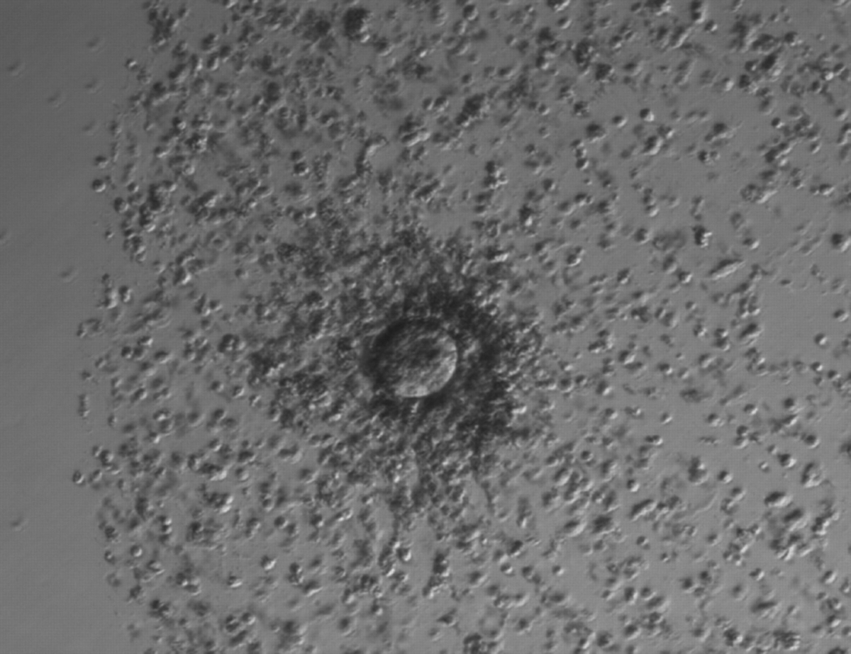
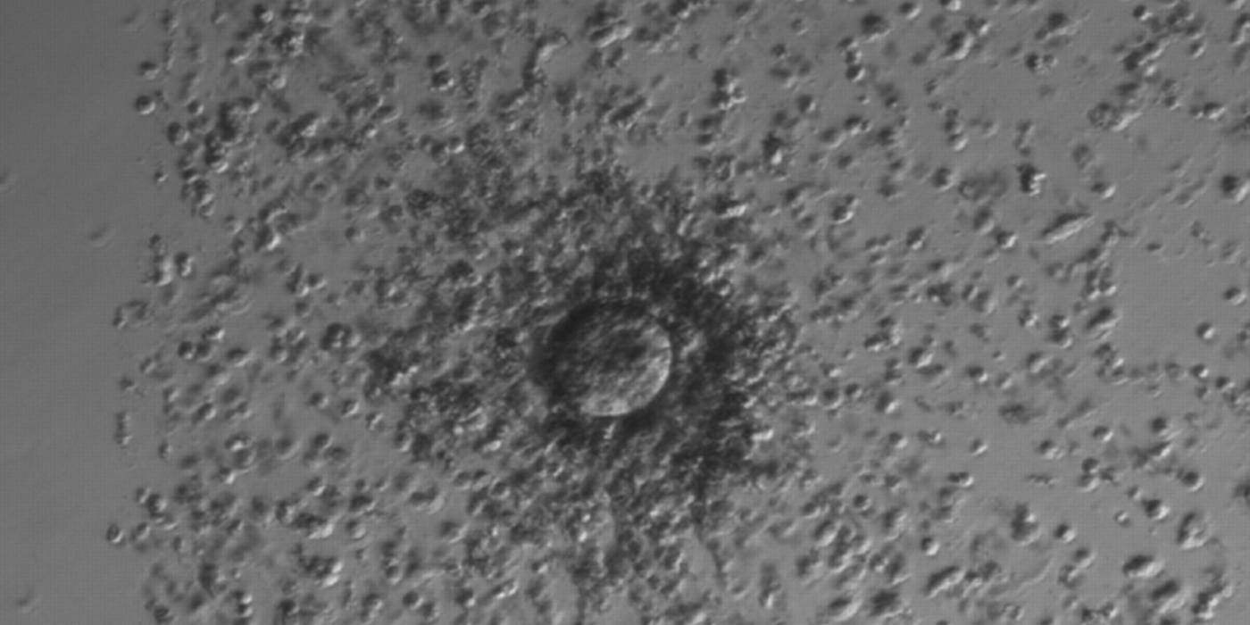
Figure 5. A cumulus oocyte complex at low magnification. The oocyte is surrounded by an expanded cumulus–corona cell complex clearly showing the separation of individual CCs due to the accumulation of hyaluronic acid in the extracellular space.

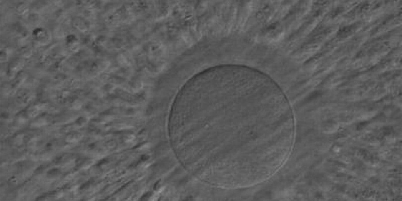
Figure 6. A cumulus–oocyte complex obtained following ovarian stimulation. An expanded cumulus corona cell complex surrounds the oocyte with the outer CCs separated from each other by extracellular matrix. The CCs immediately adjacent to the oocyte become less compact and radiate away from the ZP. The oocyte in the figure can be clearly seen through the surrounding cells at high magnification and no polar body can be seen in the PVS despite the mature status of the cumulus–corona cells.
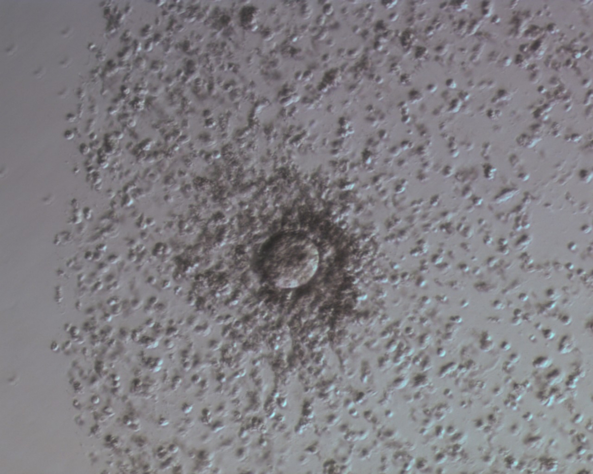
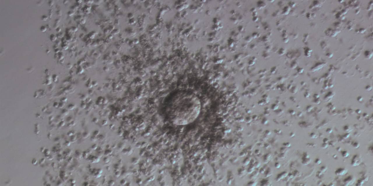
Figure 7
Denudation sequences of a mature oocyte.
a) Cumulus–corona oocyte complex before the denudation process with compact, non-radiating CCs. (100× magnification).
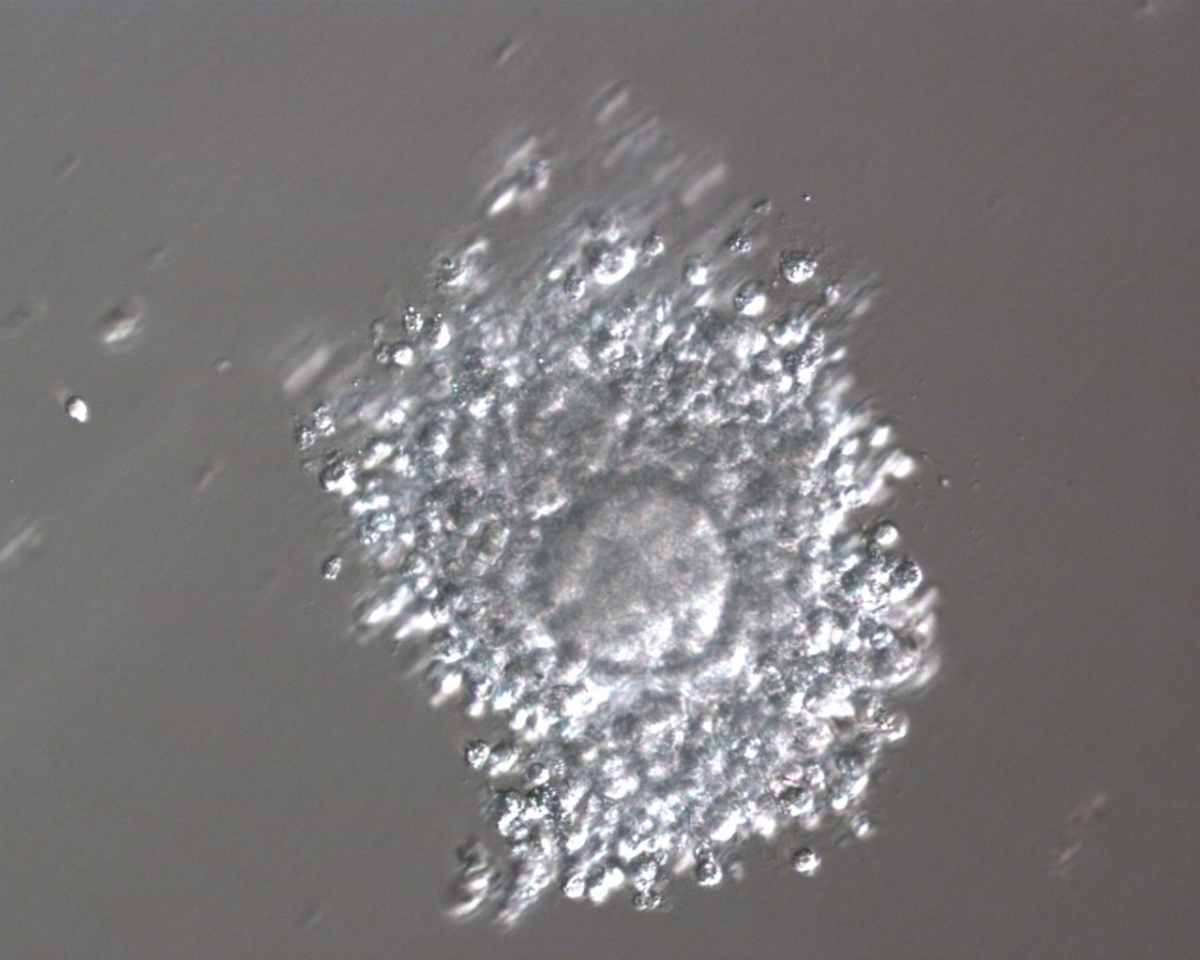
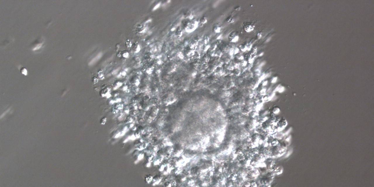
Figure 7
Denudation sequences of a mature oocyte.
b) Oocyte surrounded by corona cells during hyaluronidase treatment (200× magnification).

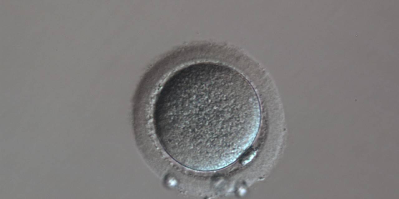
Figure 7
Denudation sequences of a mature oocyte.
c) Denuded oocyte after mechanical stripping, a visible polar body is present in the PVS (200× magnification).
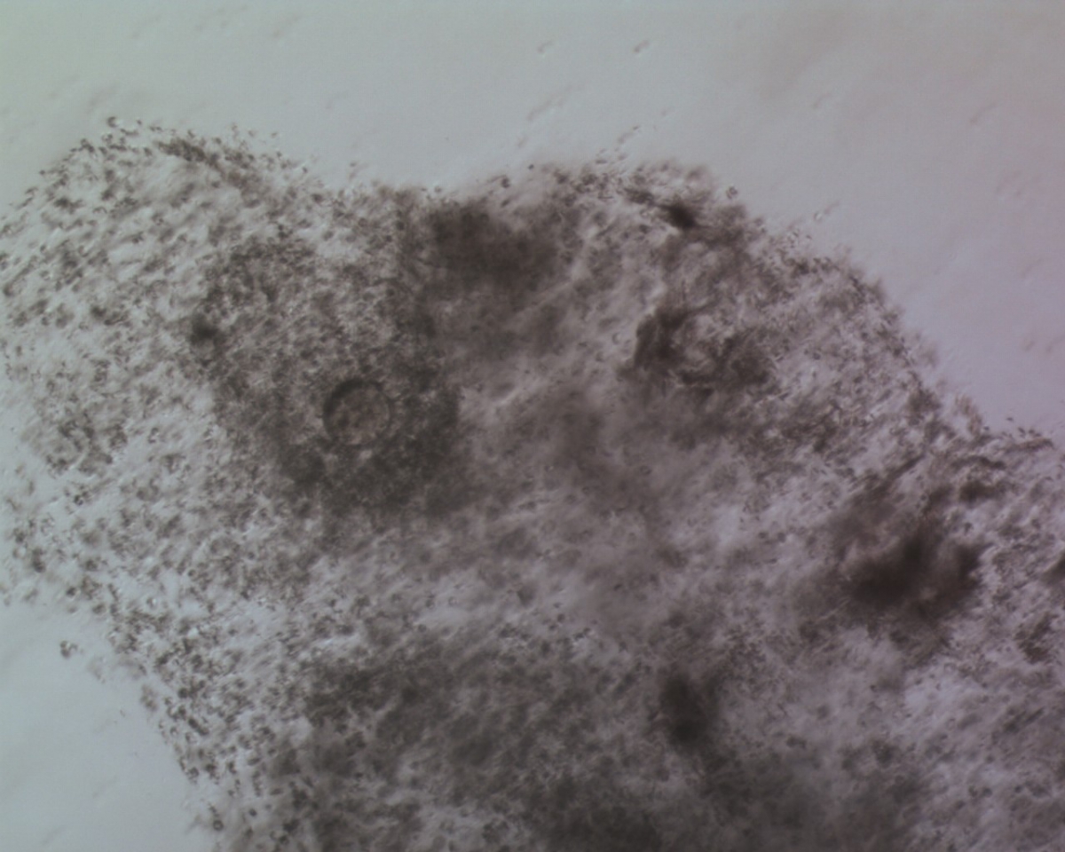
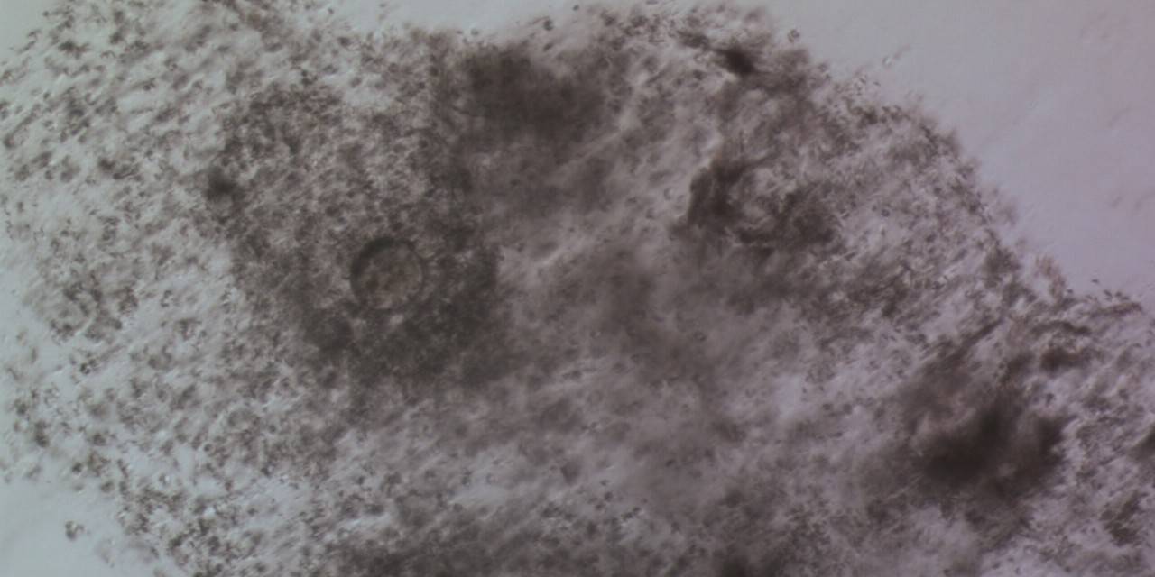
Figure 8
Denudation sequences of a mature oocyte
a) Cumulus–corona oocyte complex before the denudation process (100× magnification). CCs are abundant.
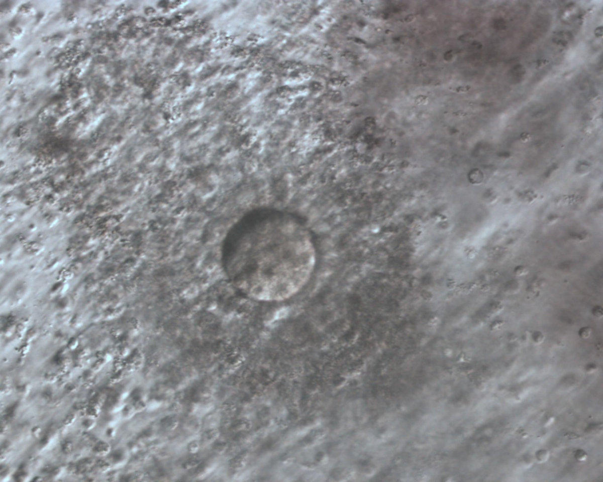
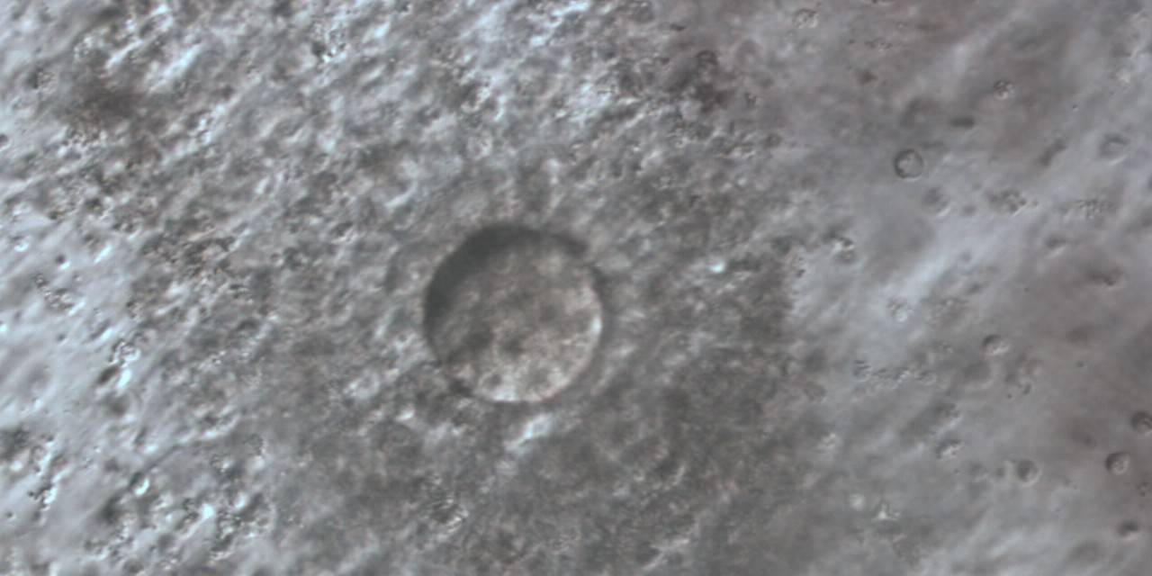
Figure 8
Denudation sequences of a mature oocyte.
b) Oocyte surrounded by corona cells during hyaluronidase treatment (200× magnification). Many CCs are still present, but the mature oocyte is already visible with PB1 at the 7 o'clock position.
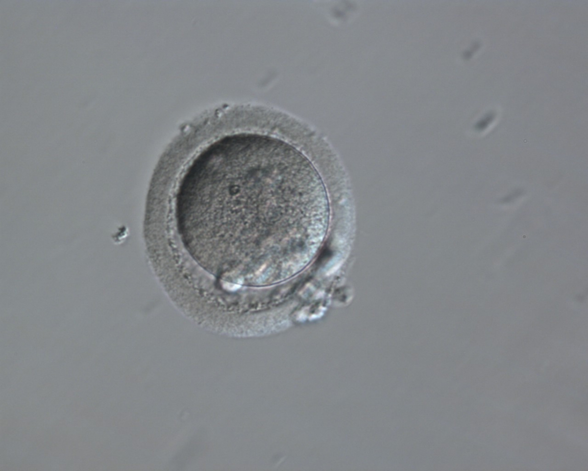
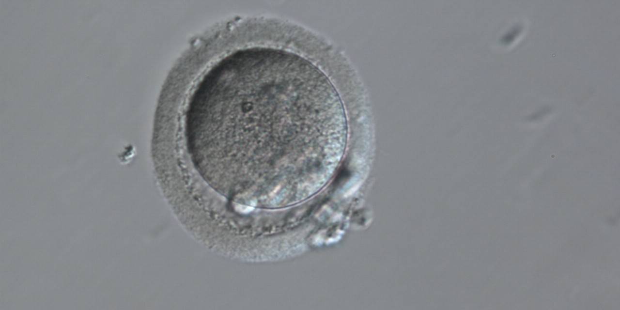
Figure 8
Denudation sequences of a mature oocyte.
c) Denuded oocyte after mechanical stripping, a visible polar body is present in the PVS (200× magnification).

Figure 9
Denudation sequences of a mature oocyte.
a) Cumulus–corona oocyte complex before the denudation process with compact, non-radiating CCs. (100× magnification).

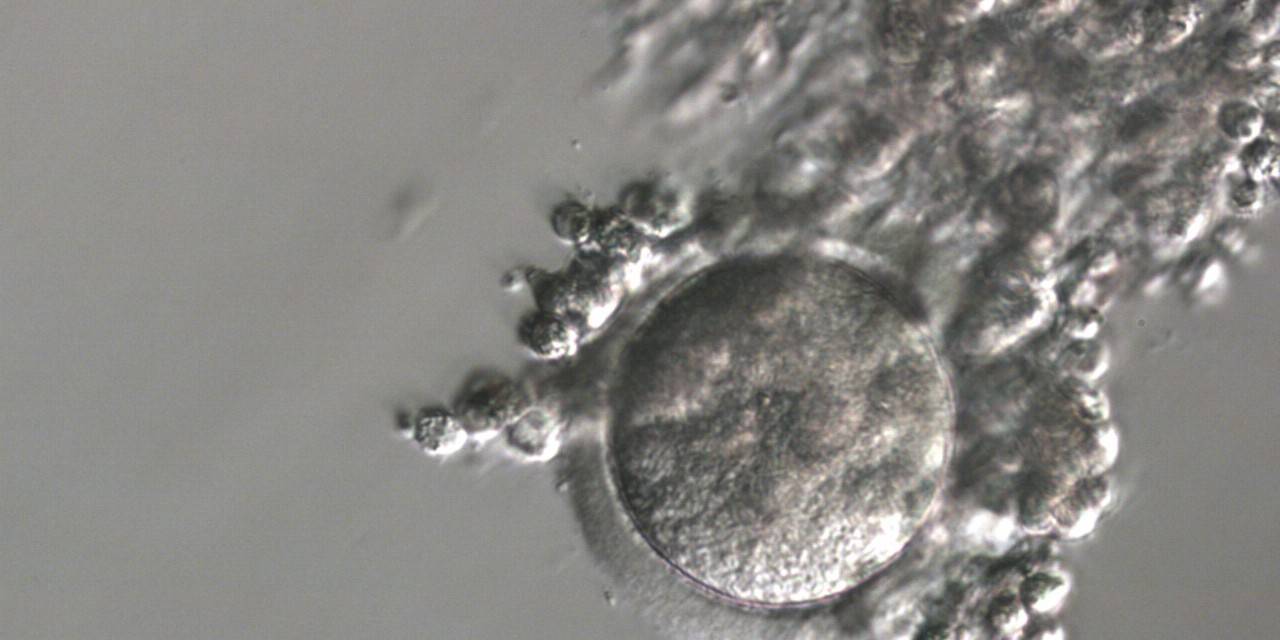
Figure 9
Denudation sequences of a mature oocyte.
b) Oocyte surrounded by corona cells during hyaluronidase treatment (200× magnification).
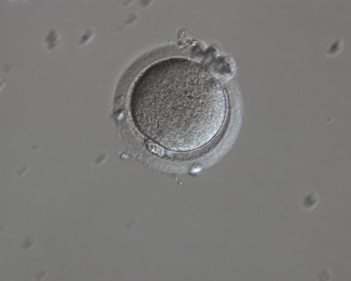
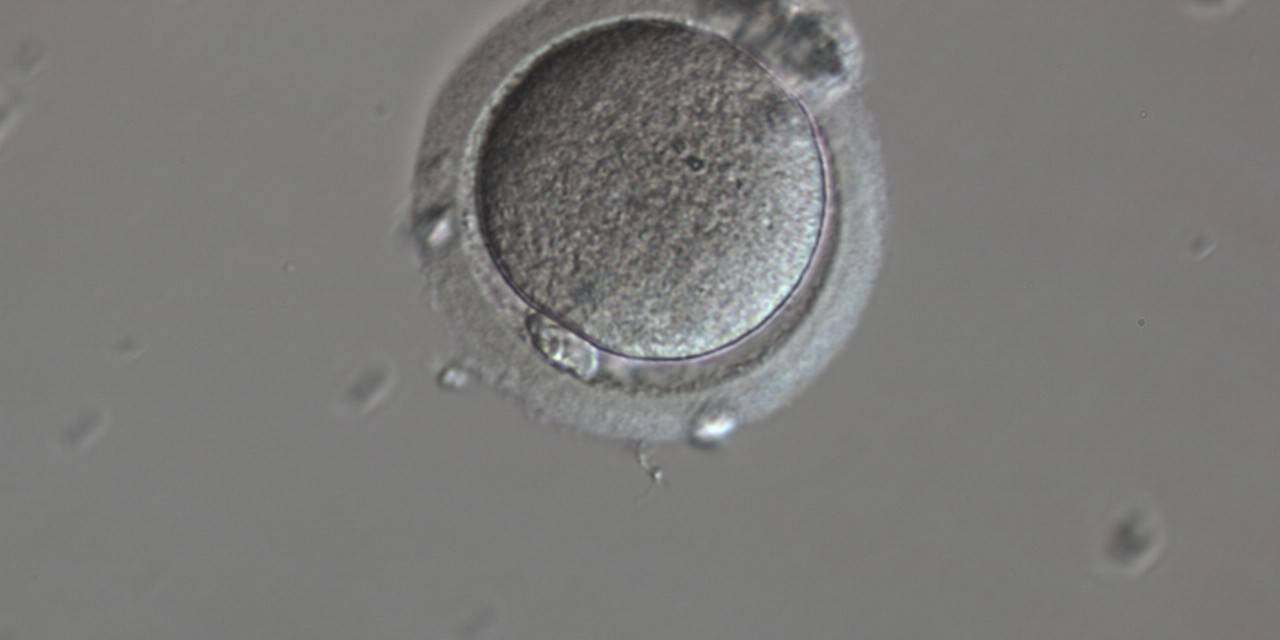
Figure 9
Denudation sequences of a mature oocyte.
c) Denuded oocyte after mechanical stripping, a visible polar body is present in the PVS (200× magnification).
The Alpha-ESHRE consensus document states that, although there is little corroborated evidence to support a correlation between the appearance of the COC and embryo developmental competence, a binary score (0 or 1) with a ‘good’ COC (score 1) defined as having an expanded cumulus and a radiating corona should be documented (Alpha Scientists in Reproductive Medicine and ESHRE Special Interest Group of Embryology, 2011).
The bidirectional communication between the oocyte and CCs, crucial for the acquisition of oocyte competence (Gilchrist et al., 2008), might perhaps be investigated in the future, through non-invasive analysis of CCs (pattern of gene expression or protein synthesis), offering new biomarkers of oocyte quality, compensating for the inadequacy of the COC morphological assessment (Feuerstein et al., 2007; Ouandaogo et al., 2011).
Article references:
Albertini DF, Combelles CM, Benecchi E, Carabatsos MJ. Cellular basis for paracrine regulation of ovarian follicle development. Reproduction 2001;121:647-653.
Abstract
Alpha Scientists in Reproductive medicine and ESHRE Special Interest Group of Embryology. The Istanbul consensus workshop on embryo assessment: proceedings of an expert meeting. Hum Reprod 2011;26:1270-1283.
Abstract/FREE Full Text
Feuerstein P, Cadoret V, Dalbies-Tran R, Guerif F, Bidault R, Royere D. Gene expression in human cumulus cells: one approach to oocyte competence. Hum Reprod 2007;22:3069-3077.
Abstract/FREE Full Text
Gilchrist RB, Lane M, Thompson JG. Oocyte-secreted factors: regulators of cumulus cell function and oocyte quality. Hum Reprod Update 2008;14:159-177.
Abstract/FREE Full Text
Laufer N, Tarlatzis BC, DeCherney AH, Masters JT, Haseltine FP, MacLusky N, Naftolin F. Asynchrony between human cumulus-corona cell complex and oocyte maturation after human menopausal gonadotropin treatment for in vitro fertilization. Fertil Steril 1984;42:366-372.
Medline | Web of Science | Google Scholar
Ouandaogo ZG, Haouzi D, Assou S, Dechaud H, Kadoch IJ, De Vos J, Hamamah S. Human cumulus cells molecular signature in relation to oocyte nuclear maturity stage. PLoSOne 2011;6:e27179.
Google Scholar
