C. Blastomere size
"STAGE SPECIFIC" VERSUS "NON-STAGE SPECIFIC"
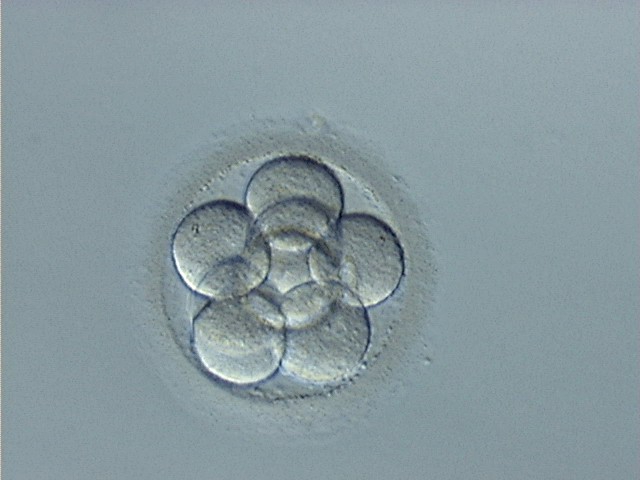
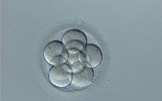
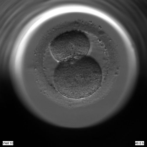
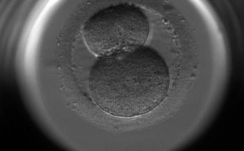
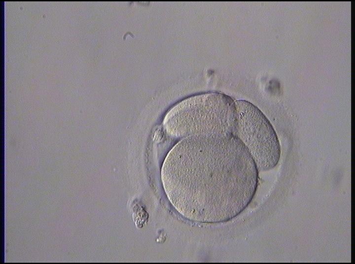
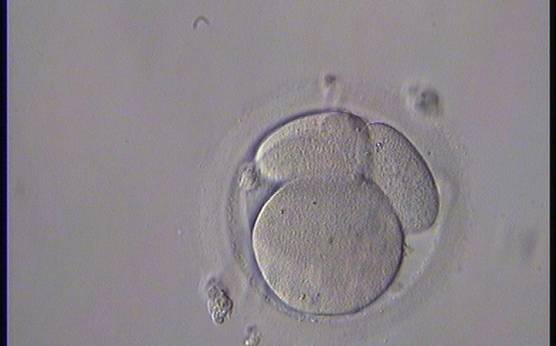
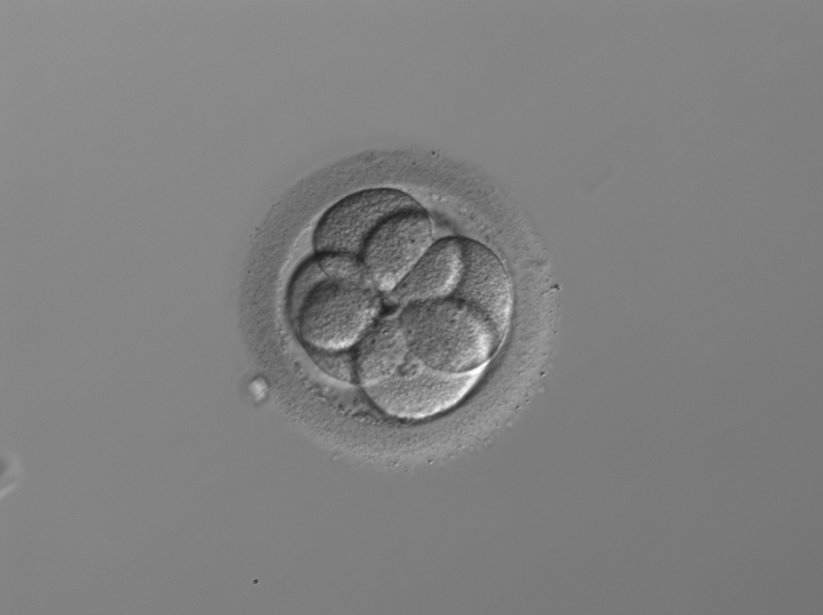
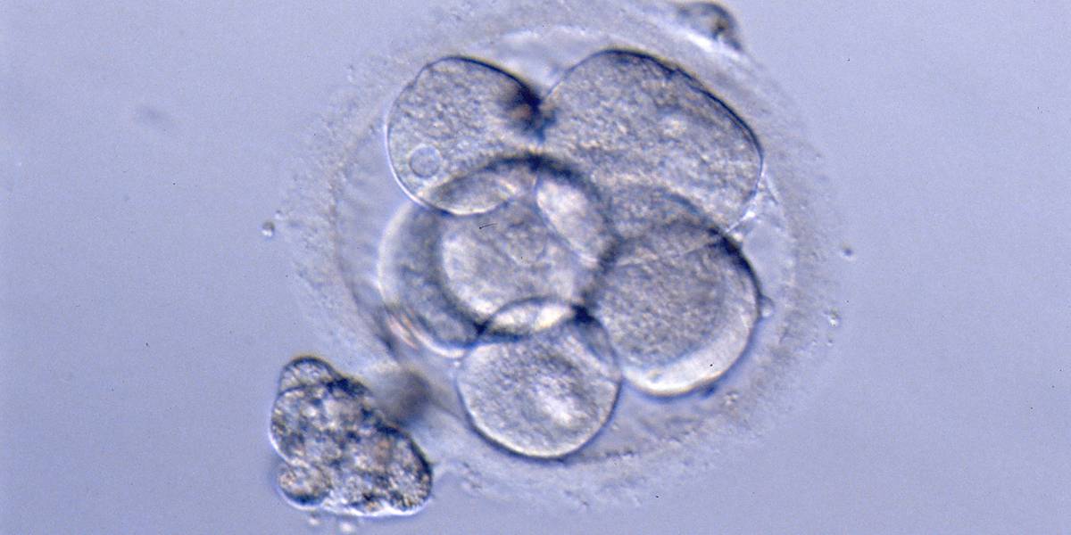
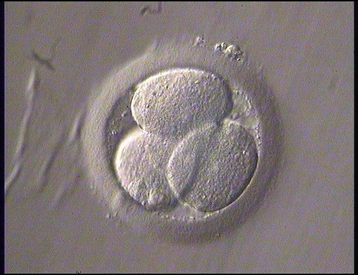
It has been shown that a high degree of regularity in the blastomere size in embryos on Day 2 is related to increased pregnancy outcome following assisted reproduction treatments (Giorgetti et al., 1995; Ziebe et al., 1997; Hardarson et al., 2001; Holte et al., 2007). Uneven cleavage, i.e. a cell cleaving into two unequal sized cells, may result in an uneven distribution of cytoplasmic molecules, e.g. proteins and mRNAs, and has been shown to be correlated with a higher incidence of multinucleation and aneuploidy (Hardarson et al., 2001; Magli et al., 2001).
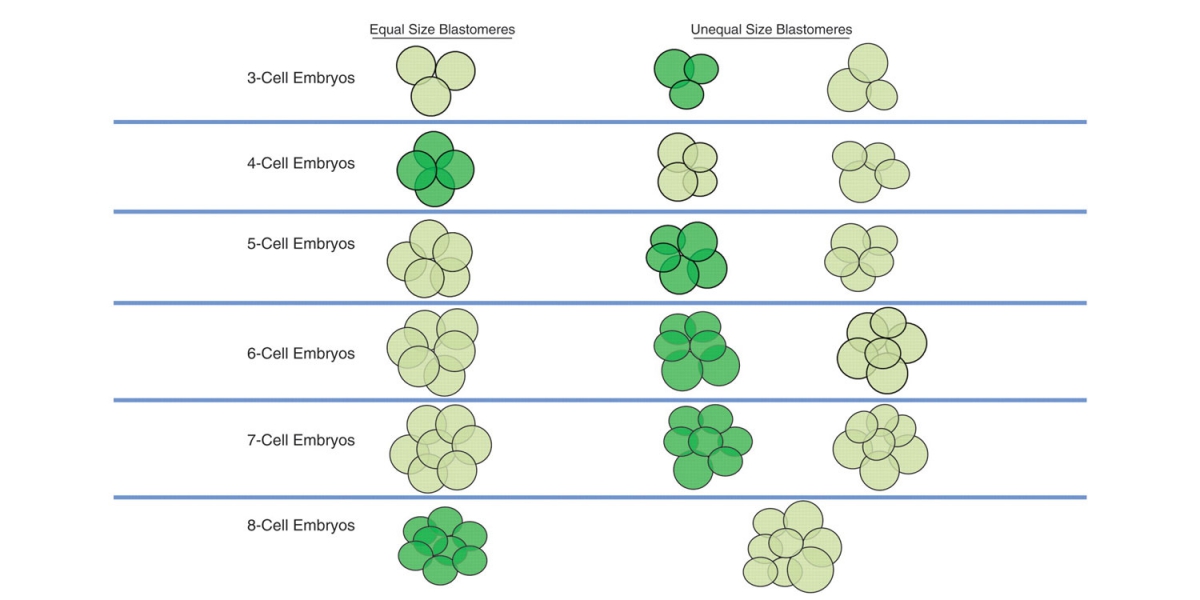
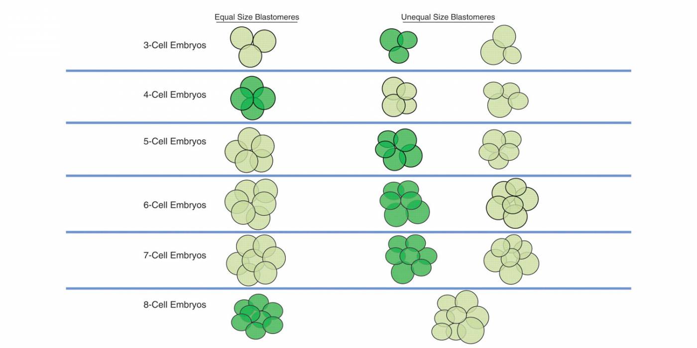
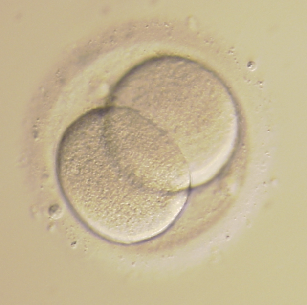
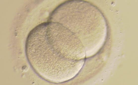
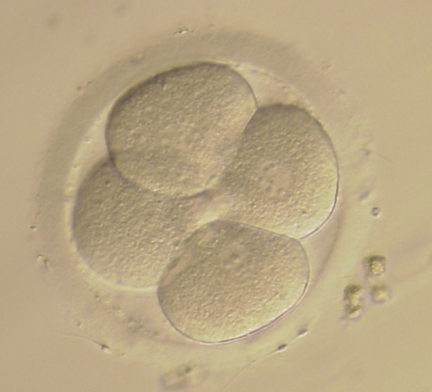
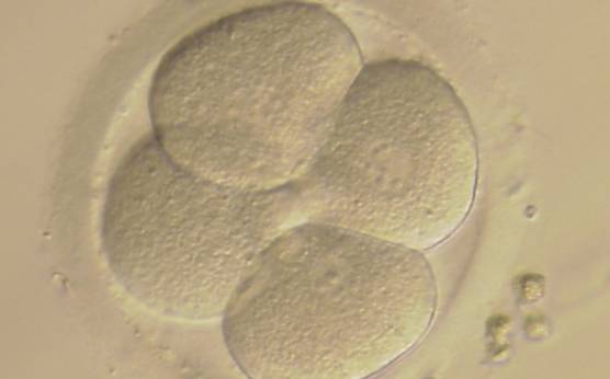
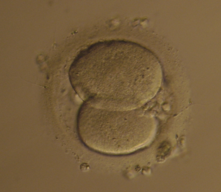
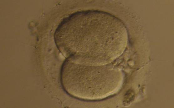
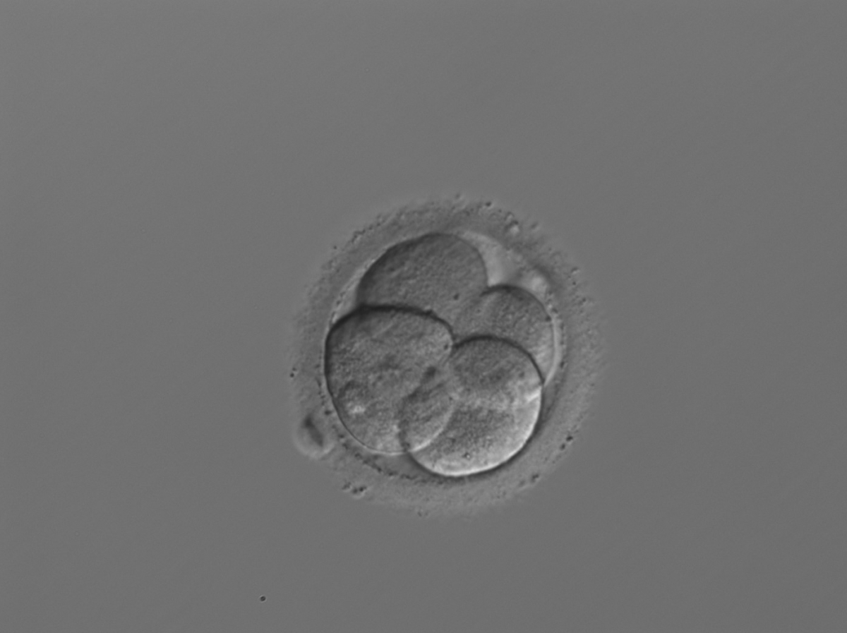
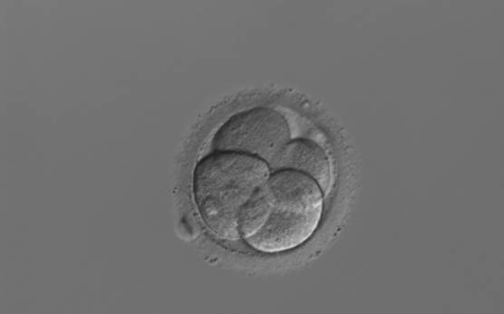
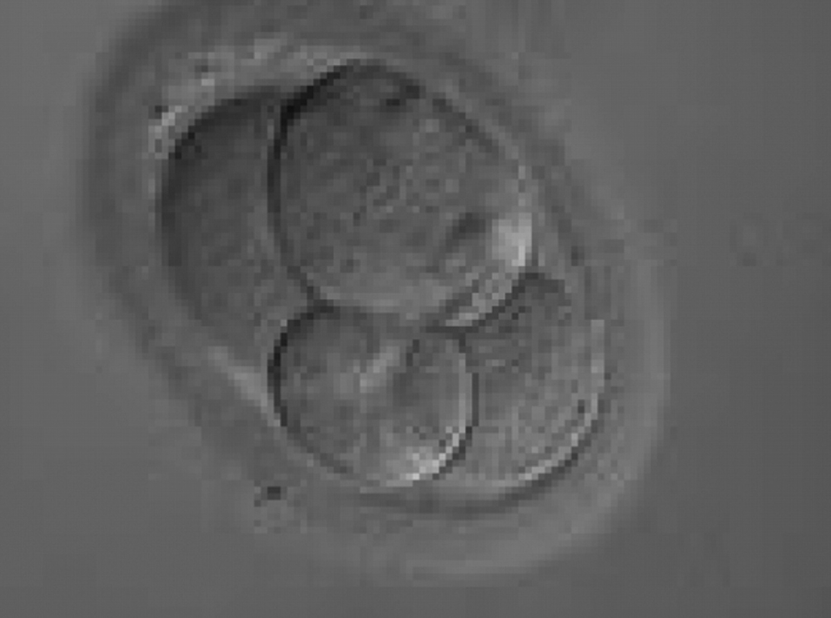
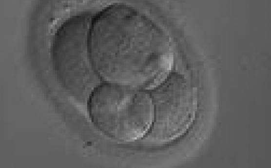
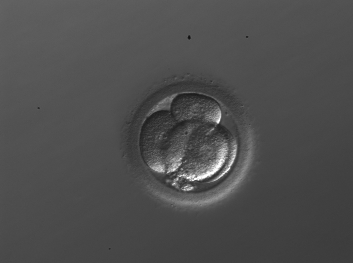
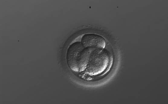
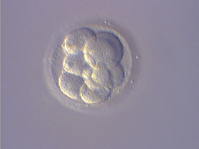
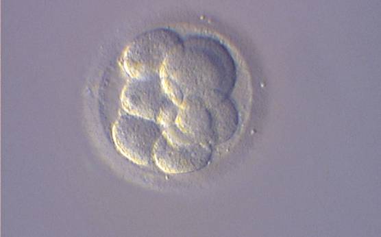
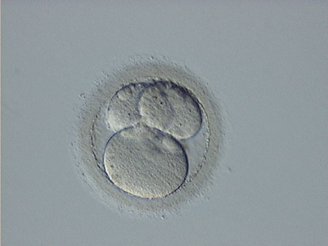
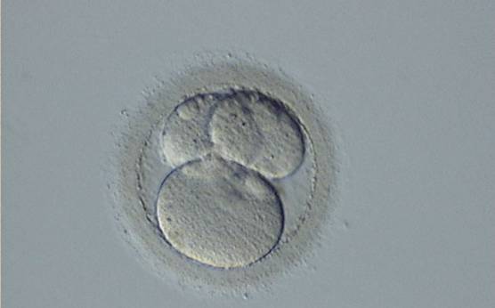
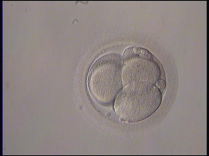
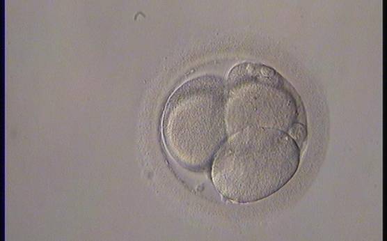
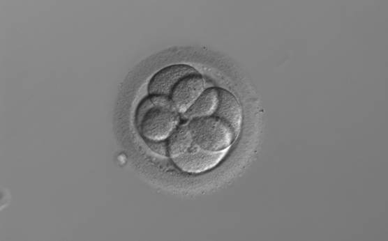
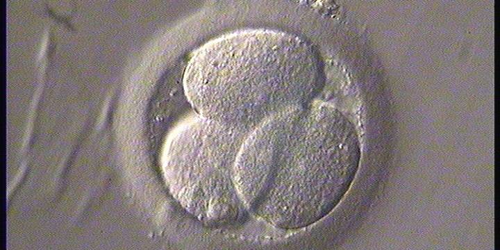
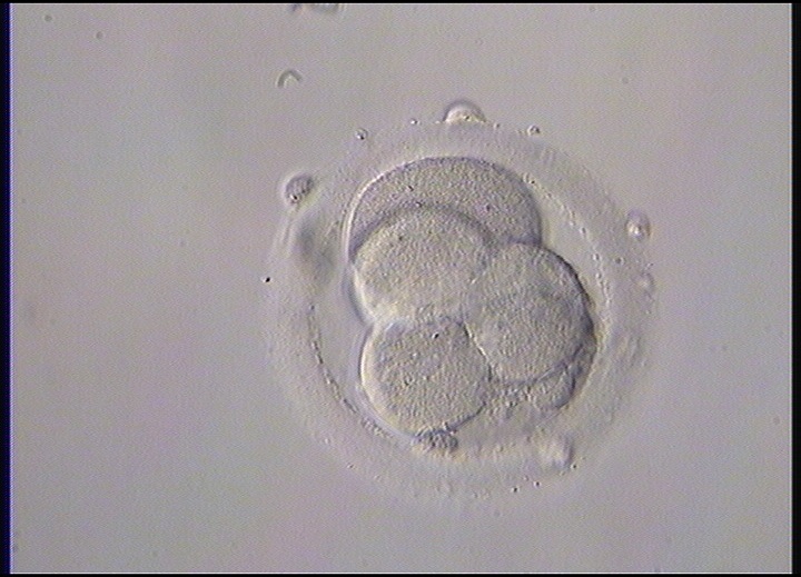
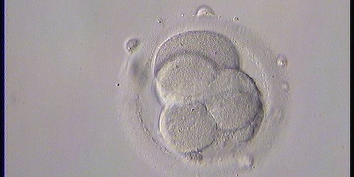
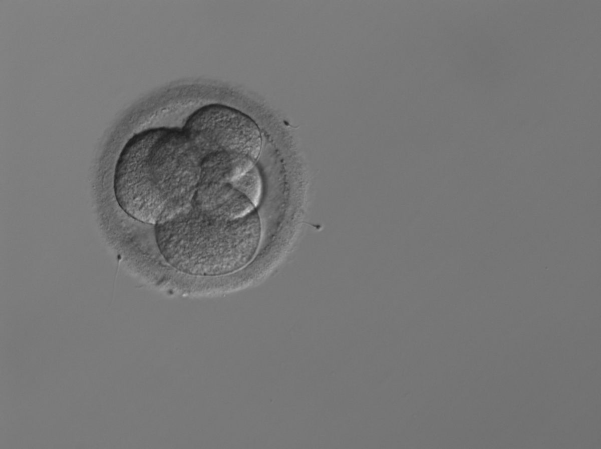
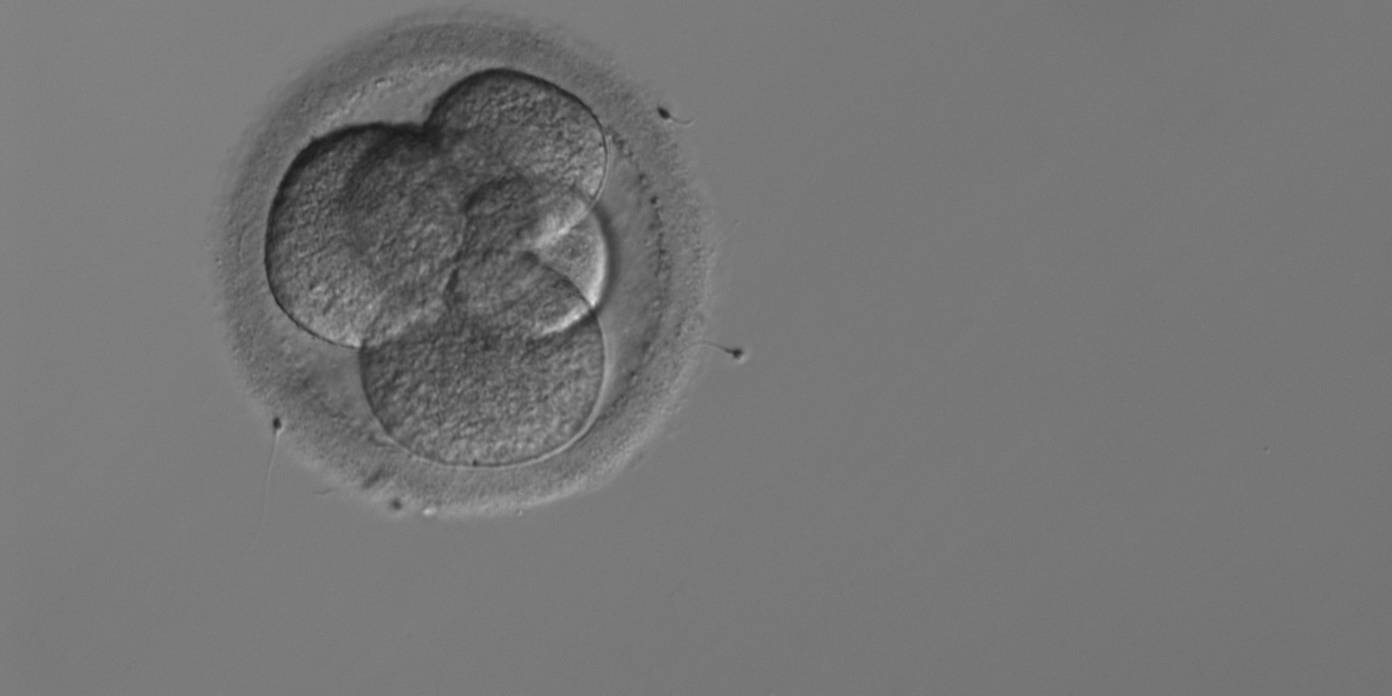
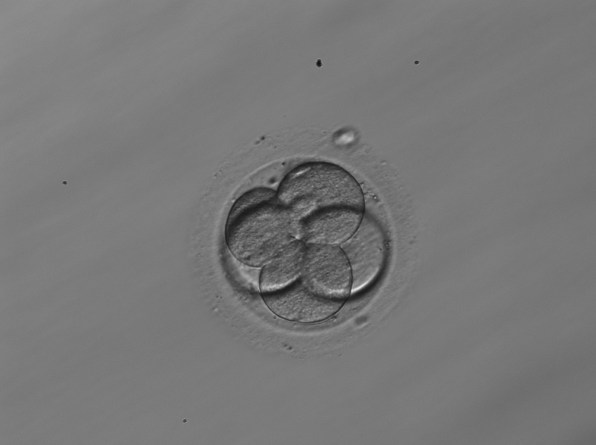
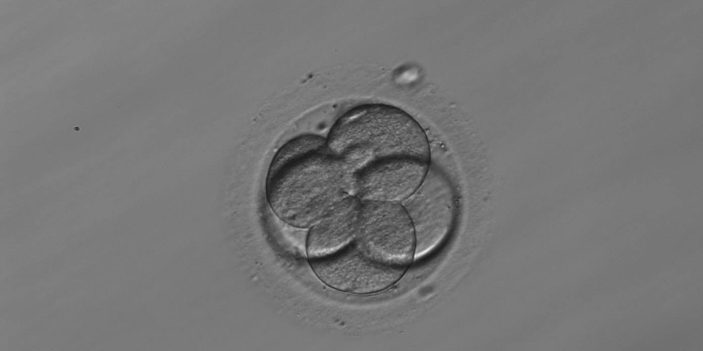
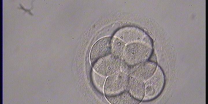
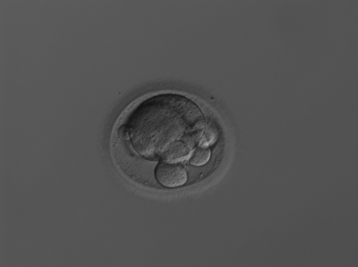
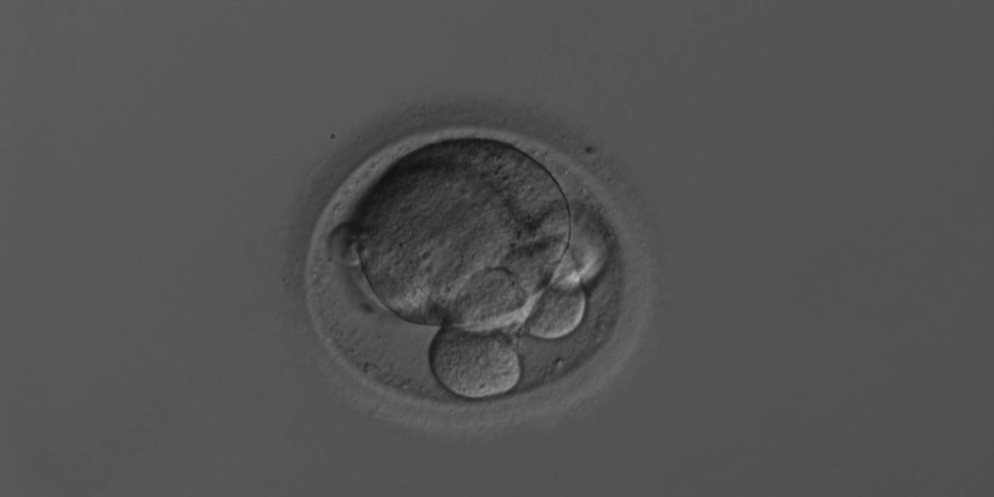
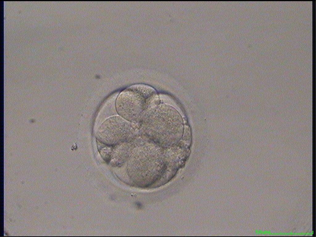
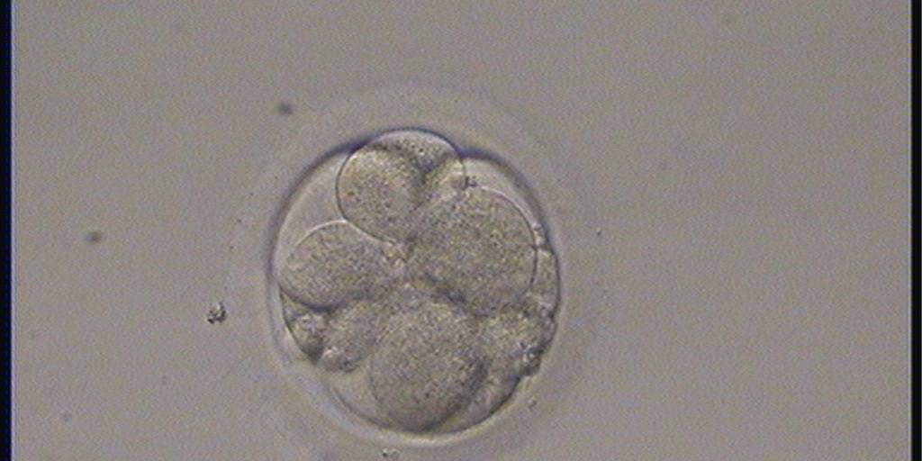
The relative blastomere size in the embryo is dependent on both the cleavage stage and the regularity of each cleavage division (Diagrams 1 and 2). The blastomeres of 2-, 4- and 8-cell embryos should be equal (stage-specific embryos, Figs 243–245) rather than unequal in size (non-stage-specific embryos, Figs 246–252). In contrast, blastomeres of embryos with cell numbers other than 2, 4 and 8 should have different sizes as there is an asynchrony in the division of one or more blastomeres (Figs 253–256). A 3-cell embryo should preferably have one large and two small blastomeres (Fig. 253); a 5-cell embryo, three large and two smaller blastomeres (Fig. 254); a 6-cell embryo, two large and four smaller blastomeres (Fig. 255) and a 7-cell embryo, one large and six smaller blastomeres (Fig. 256). These embryos are thereby also considered to be stage specific. However, a 4-cell embryo with one or two blastomers much larger than the others (Figs 248–251), a 3-cell embryo with all blastomeres even in size (Fig. 257), a 5-cell embryo with two large and three smaller blastomeres (Figs 258 and 259) or one small and four larger blastomeres (Fig. 260), a 6-cell embryo with all blastomeres even in size (Fig. 261) or extremely different in size (Fig. 262) and a 7-cell embryo with three large and four smaller blastomeres (Fig. 263) would not be considered to have normal blastomere sizes in relation to cell numbers and are therefore not considered to be stage specific (for further clarification see Diagrams 1 and 2).
Article references:
Giorgetti C, Terriou P, Auquier P, Hans E, Spach JL, Salzmann J, Roulier R. Embryo score to predict implantation after in-vitro fertilization: based on 957 single embryo transfers. Hum Reprod 1995;10:2427-2431.
Abstract/FREE Full Text
Hardarson T, Hanson C, Sjögren A, Lundin K. Human embryos with unevenly sized blastomeres have lower pregnancy and implantation rates: indications for aneuploidy and multinucleation. Hum Reprod 2001;16:313-318.
Abstract/FREE Full Text
Holte J, Berglund L, Milton K, Garello C, Gennarelli G, Revelli A, Bergh T. Construction of an evidence-based integrated morphology cleavage embryo score for implantation potential of embryos scored and transferred on day 2 after oocyte retrieval. Hum Reprod 2007;22:548-557.
Abstract/FREE Full Text
Magli MC, Gianaroli L, Ferraretti AP. Chromosomal abnormalities in embryos. Mol Cell Endocrinol 2001;183:29-34.
CrossRef | Medline | Web of Science | Google Scholar
Ziebe S, Petersen K, Lindenberg S, Andersen AG, Gabrielsen A, Andersen AN. Embryo morphology or cleavage stage: how to select the best embryos for transfer after in-vitro fertilization. Hum Reprod 1997;12:1545-1549.
Abstract/FREE Full Text

