E. Extracytoplasmatic features
By definition, extracytoplasmic anomalies of the egg include all dysmorphisms related to the ZP, PVS and the polar body of the mature oocyte.
E.1 Zona pellucida
Any alterations in ZP appearance could be caused by secretion and patterning problems of the glycoprotein matrix (Shen et al., 2005). Since apparent changes in thickness (Figs 61–64) or complete absence of the ZP are extremely rare (Stanger et al., 2001), more subtle changes in the three-dimensional structure are most frequently observed. Since the inner layer of the zona is highly ordered, it can clearly be visualized using polarized light (Figs 65–68; Pelletier et al., 2004). Embryos with a good prognosis in terms of blastocyst formation and pregnancy can be predicted when viewed using polarized light (Montag et al., 2008; Ebner et al., 2010).
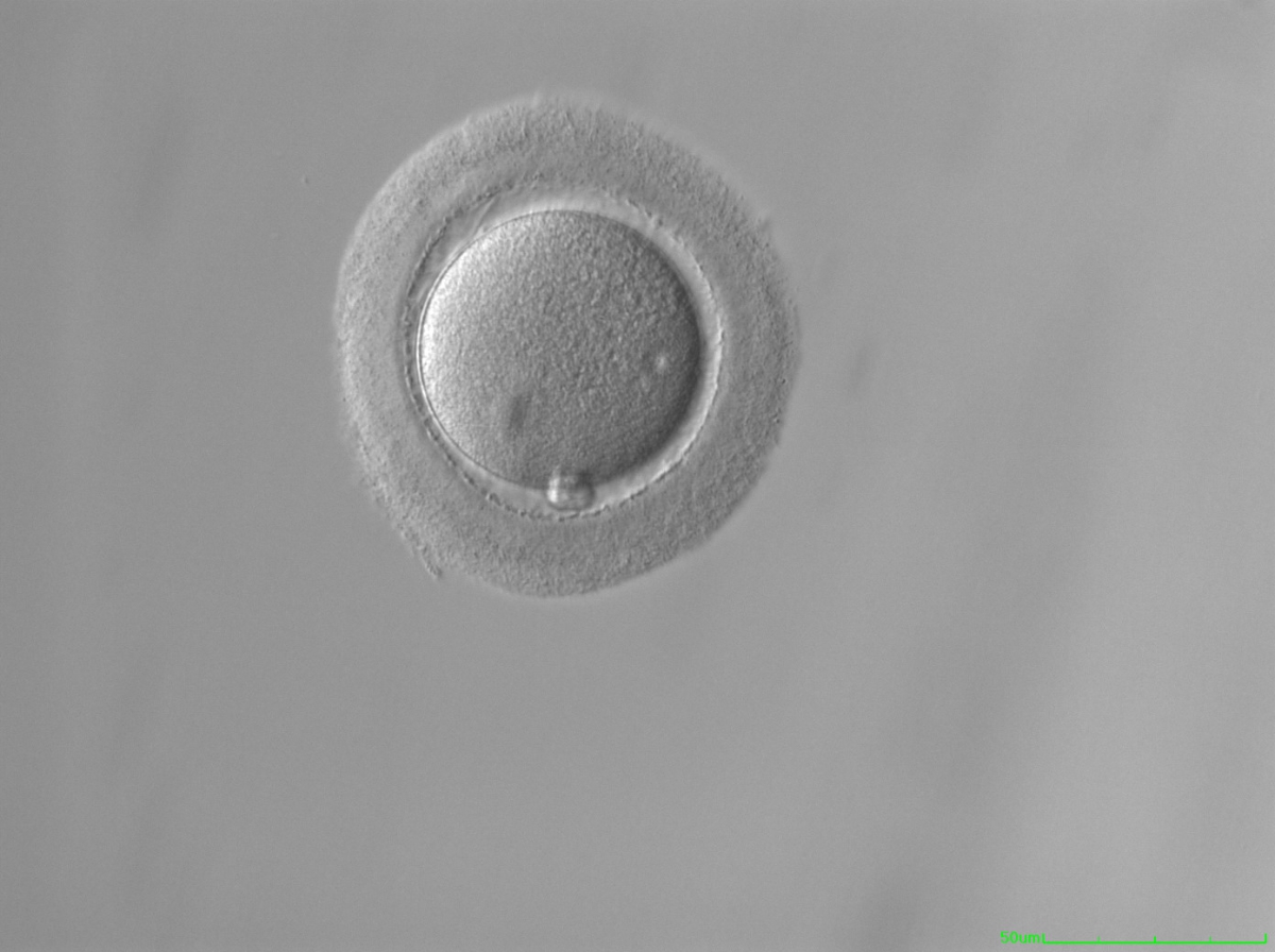
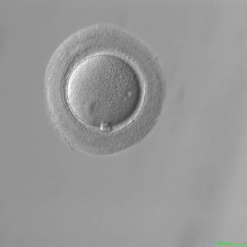
Figure 61
MII oocyte with a thick and dense ZP (200× magnification).


Figure 62
MII oocyte with a thick ZP (200× magnification).
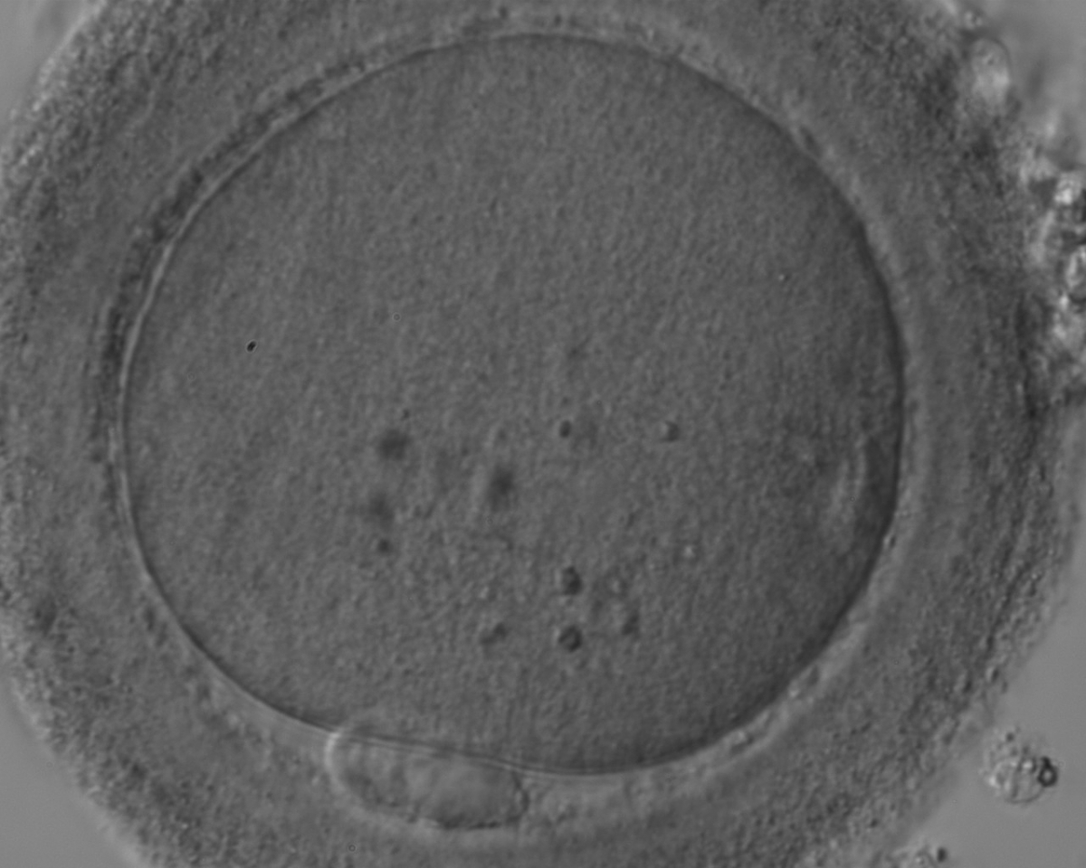
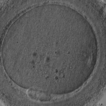
Figure 63
MII oocyte with a thick and dark ZP. Some inclusions are visible in the ooplasm.
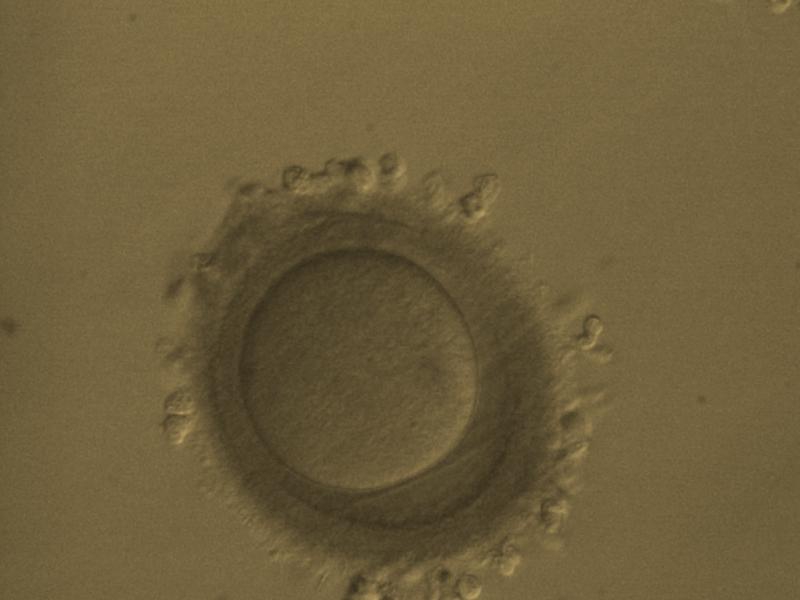
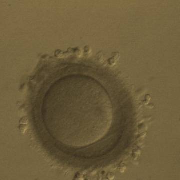
Figure 64
MII oocyte with a thick and dark ZP whose thickness is not homogeneous
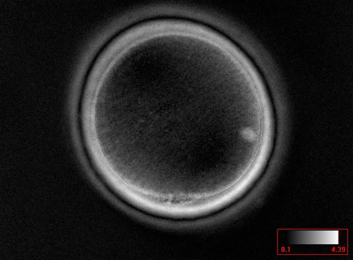
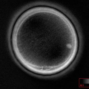
Figure 65
MII oocyte observed using polarized light microscopy with a clear birefringent ZP inner layer. The MS is dislocated from PB1 by almost 90°.
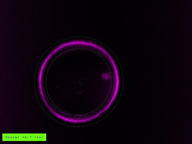
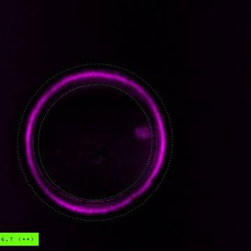
Figure 66
MII oocyte observed using polarized light microscopy with a clear birefringent ZP inner layer. The MS is also clearly visible at the 3 o'clock position in this view.
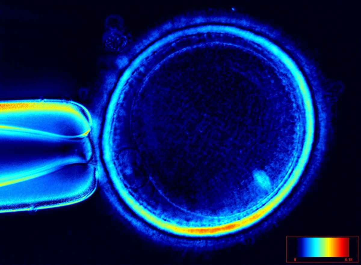
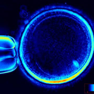
Figure 67
MII oocyte showing different degrees of ZP inner layer birefringence. The figure has a high degree of birefringence.
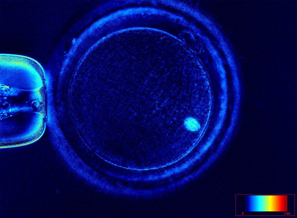
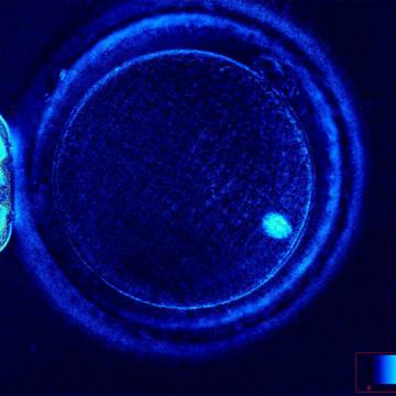
Figure 68
MII oocyte showing different degrees of ZP inner layer birefringence. The figure has a low degree of birefringence.
The degree to which discoloration of the ZP contributes to the birefringence of the outer shell is not known; however, it has been suggested that successful fertilization, embryo development and pregnancy can be achieved after transfer of embryos derived from brown zonae (Figs 63 and 64; Esfandiari et al., 2006).
Another rare finding is grossly abnormally shaped ZP with what could be either duplication of the inner layer of the ZP or a tear in the layers of the ZP creating an intrazonal space (Figs 69–71).
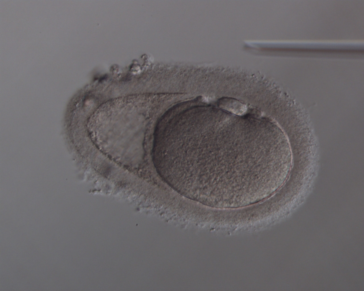
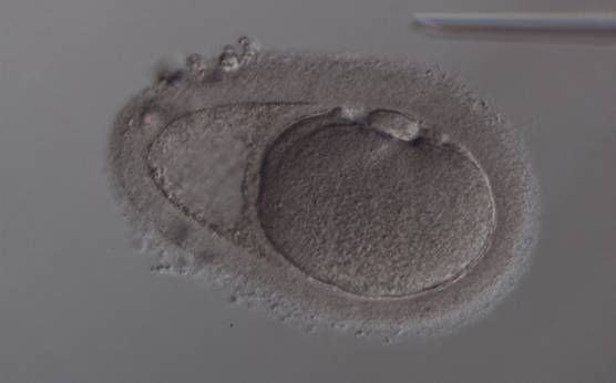
Figure 69
Oocyte with an abnormally shaped ZP and with what appears to be a duplication of, or tears in, the ZP. The oocyte has an irregular shape and cytoplasmic appearance with a large sized PB1.

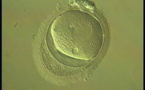
Figure 70
Oocyte with an abnormally shaped ZP and with what appears to be a duplication of, or tears in, the ZP. The oocyte is highly dysmorphic with no evident PB1 in the PVS.
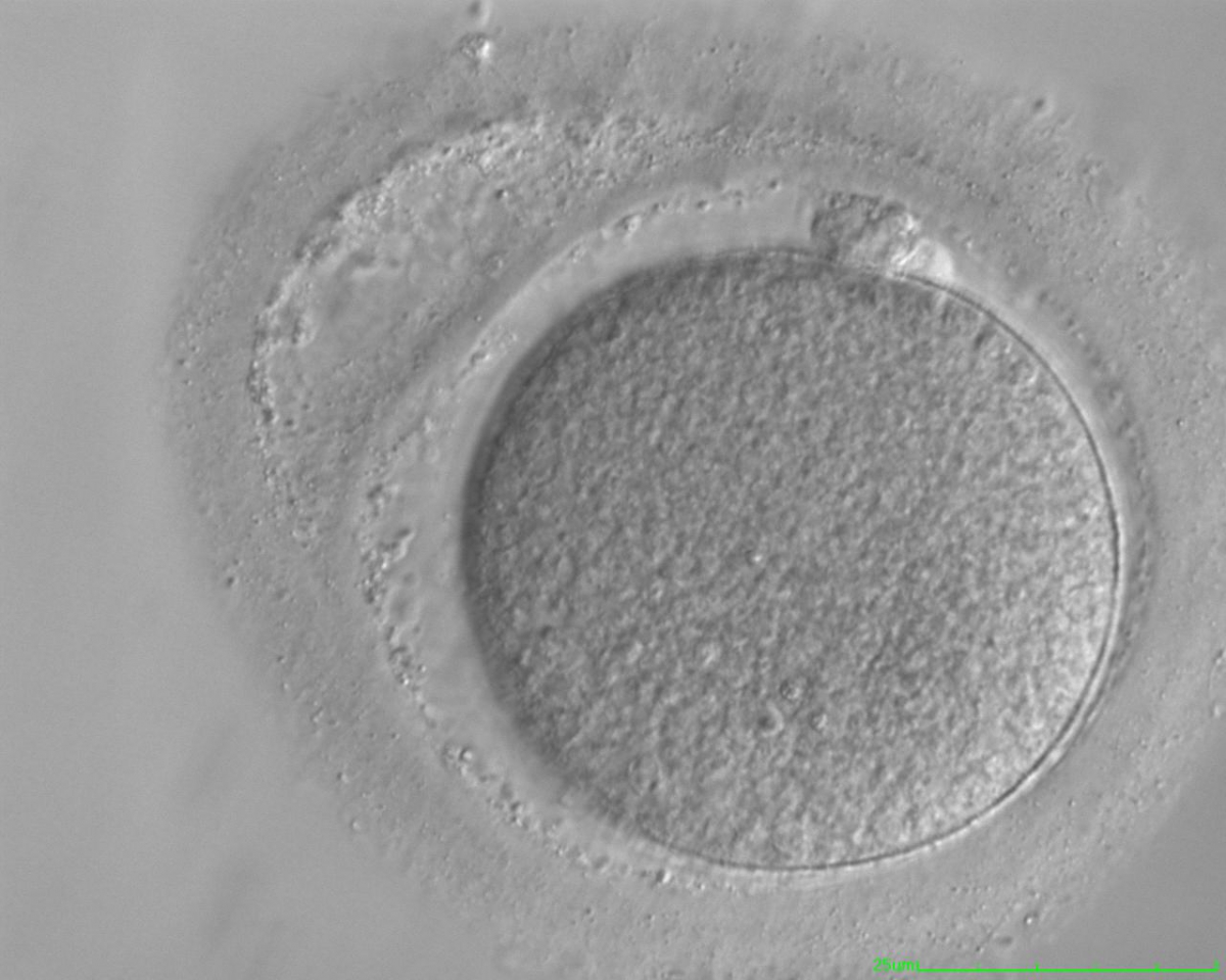
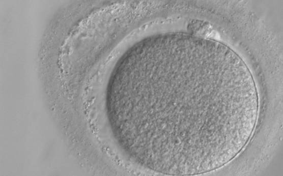
Figure 71
Oocyte with an abnormally shaped ZP and with what appears to be a duplication of, or tears in, the ZP. The oocyte has a regular shape.
E.2 Perivitelline space
Several authors have noted that approximately one-third of all ova show a large PVS, a dysmorphism that was found to be negatively correlated with fertilization rate and embryo quality (Xia, 1997; Rienzi et al., 2008). Data from the literature indicate that a large PVS (Figs 72–74) may be ascribed to over-mature eggs (Mikkelsen and Lindenberg, 2001; Miao et al., 2009). In other words, such eggs have shrunk in relation to the ZP presenting a large gap between the oocyte and surrounding zona. A large PVS would also occur if a larger portion of cytoplasm is extruded together with the haploid chromosomal set during PBI formation. This would result in a large PBI and as a consequence a large PVS.
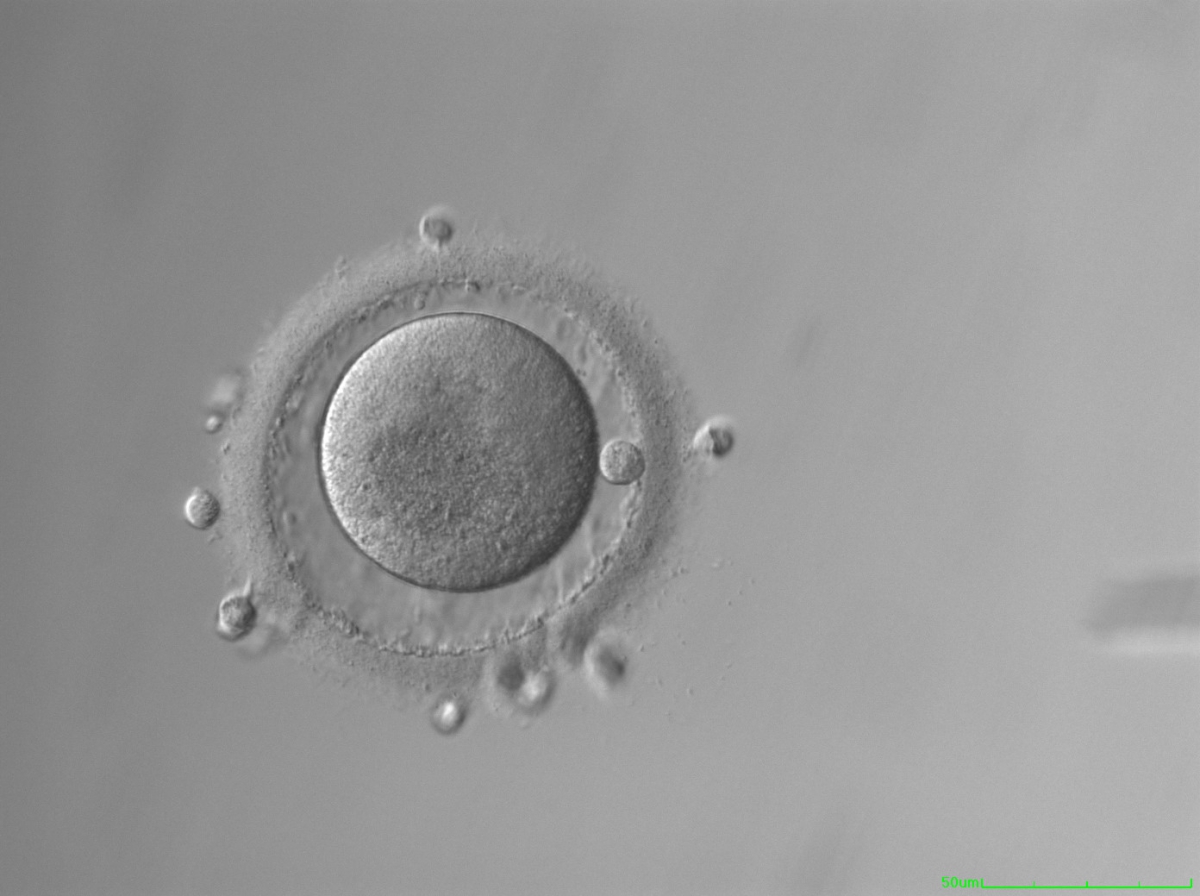
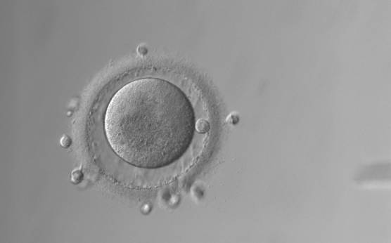
Figure 72
Oocyte with a large PVS. There is a large granular area in the cytoplasm.
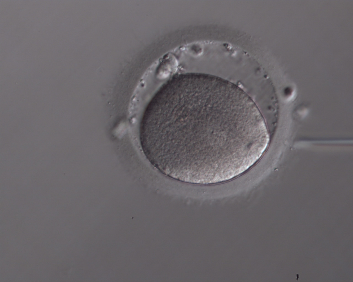
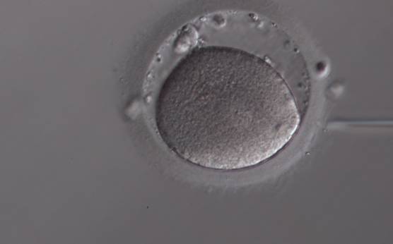
Figure 73
Oocyte with a large PVS. Several fragments are present in the PVS.
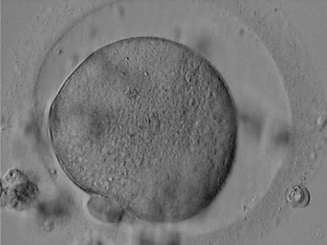

Figure 74
Oocyte with a large PVS and a granular cytoplasm.
E.3 Polar body
Generally, PBI morphology can be seen as a reflection of postovulatory age of the oocytes since this by-product of meiosis fragments with time. Nevertheless, the impact of PBI morphology on outcome is still a matter of debate. Oocytes showing an intact PBI (Fig. 75) give rise to higher rates of implantation and pregnancy (Ebner et al., 1999) probably due to an increase in blastocyst formation (Ebner et al., 2002). Apparently, the benefit of selecting oocytes according to the morphology of PBI is diluted with increasing time between ovulation induction and ICSI, since studies with different schedules could not find a relationship between morphology of PBI and subsequent ICSI outcome (Ciotti et al., 2004; De Santis et al., 2005; Fancsovits et al., 2006).
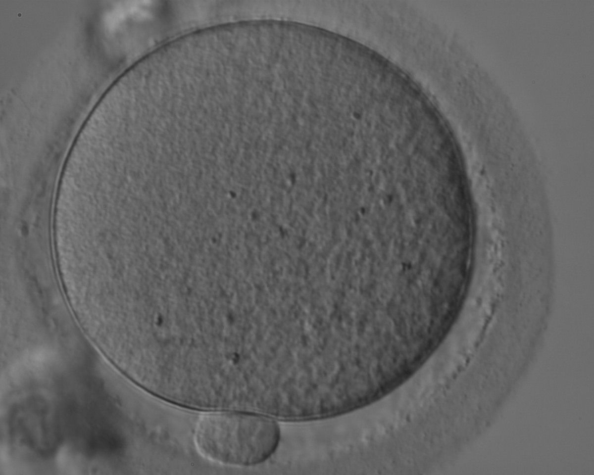
Figure 75
MII oocyte with a normal-sized PBI.
However, the fact that a large PBI (Figs 76–78) has a very poor prognosis remains unchallenged (Fancsovits et al., 2006). When large PBI's are extruded, embryos with multinucleated blastomeres are a significantly more frequent consequence than for all other PBI morphological categories (Fancsovits et al., 2006). It has been postulated that the extrusion of an abnormally large PBI is due to dislocation of the MS (Verlhac et al., 2000).
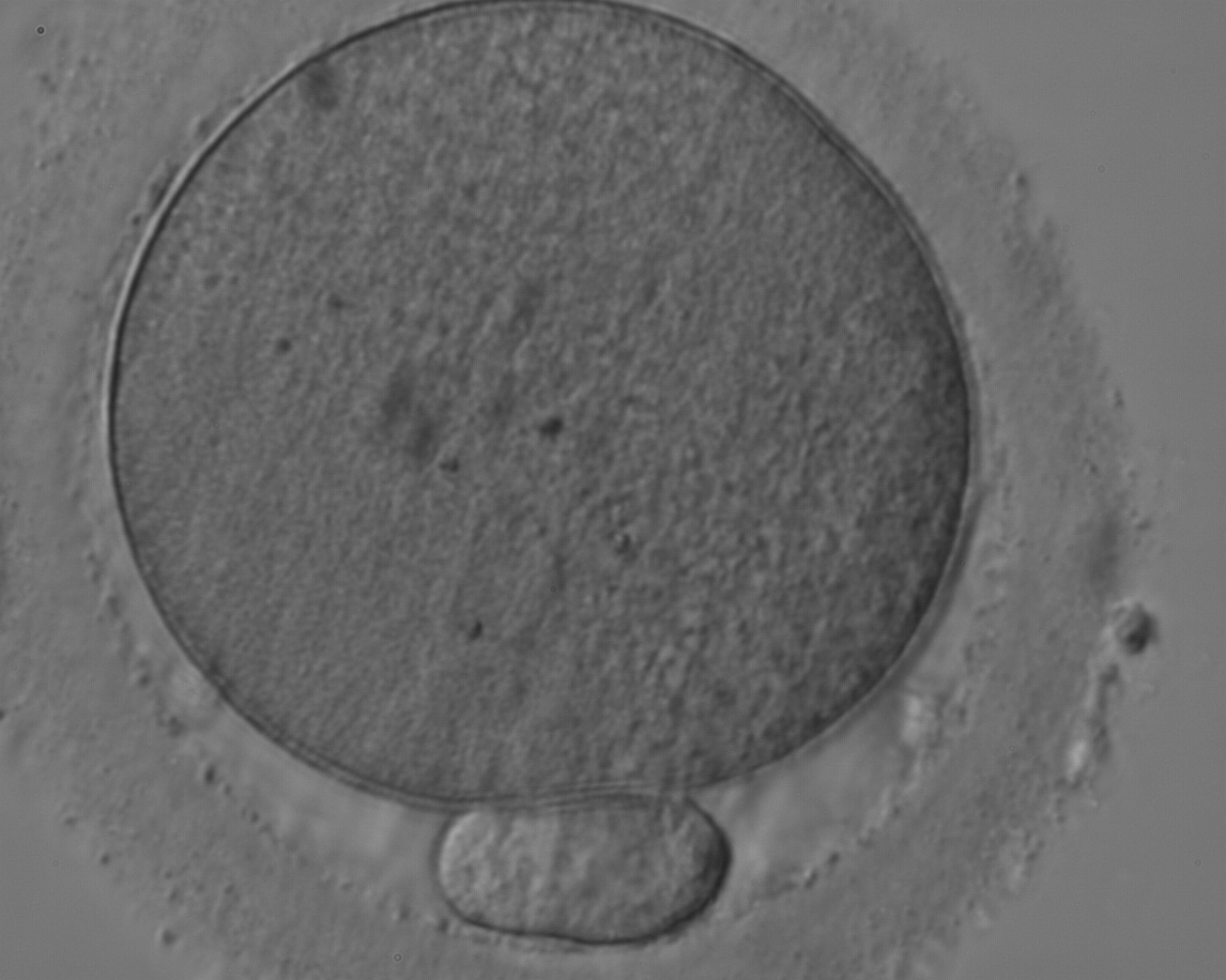
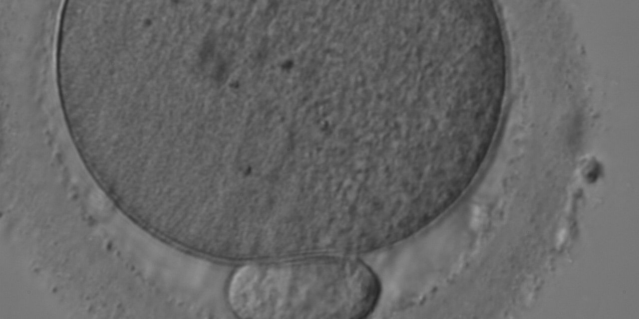
Figure 76
MII oocyte with a large PBI, 3–4 times larger than normal.
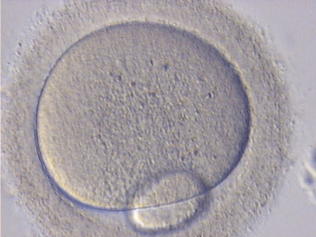
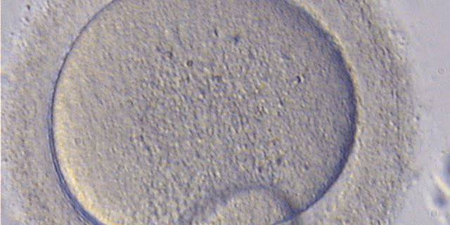
Figure 77
MII oocyte with a giant PBI, 5-6 times larger than normal.
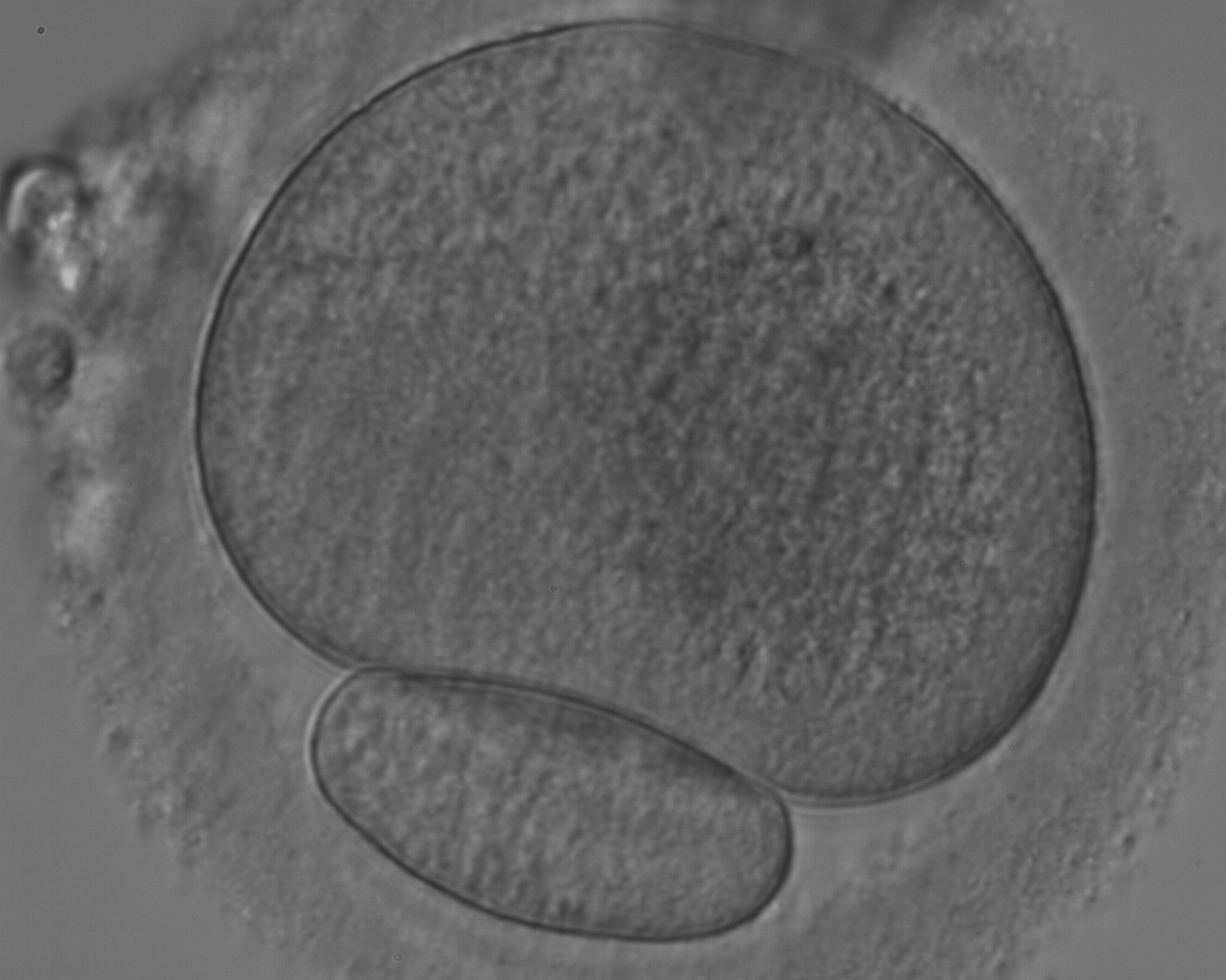
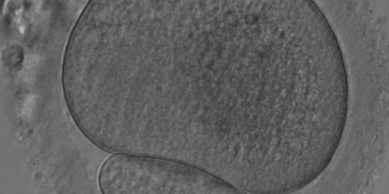
Figure 78
MII oocyte with a giant PBI.
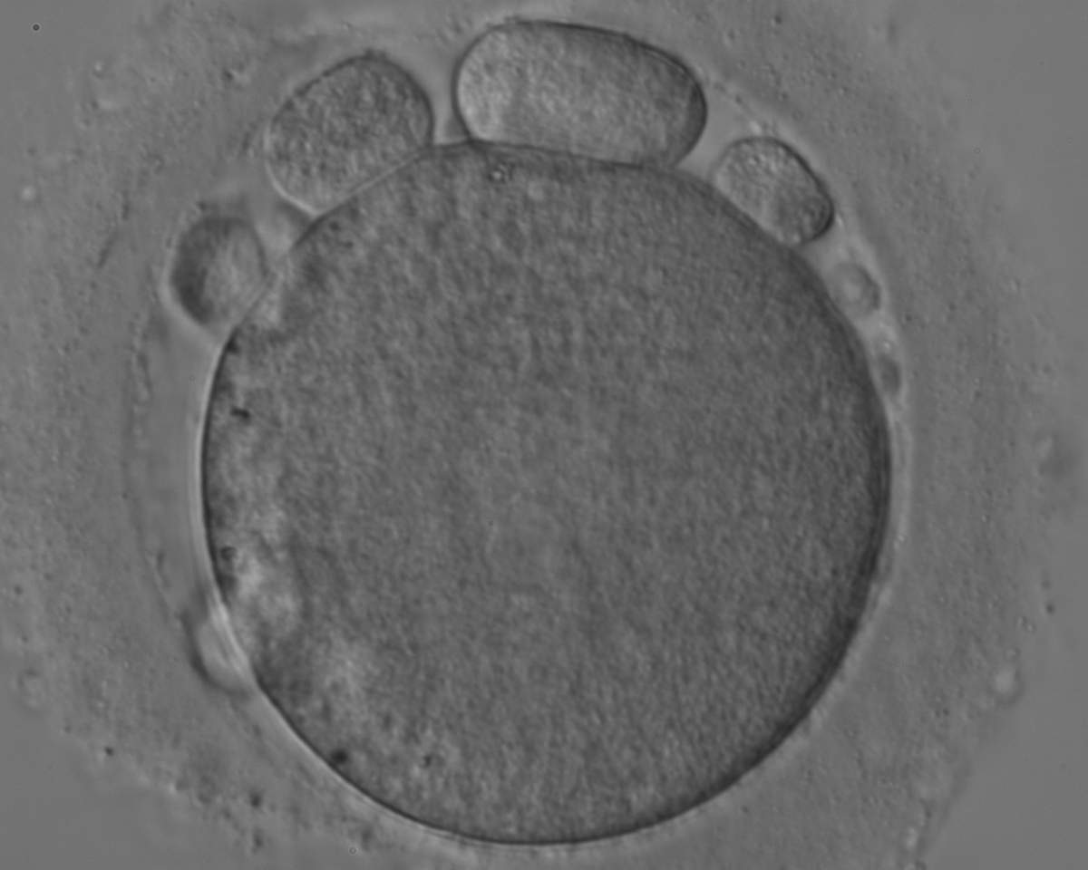
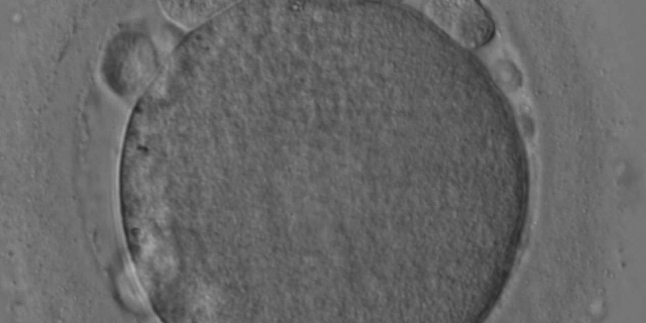
Figure 79
MII oocyte with large fragments and a large PBI in the PVS.
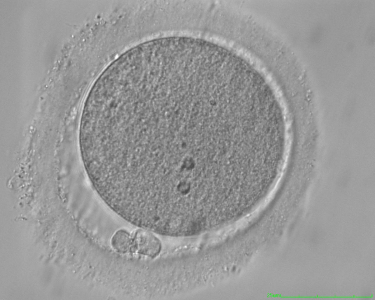
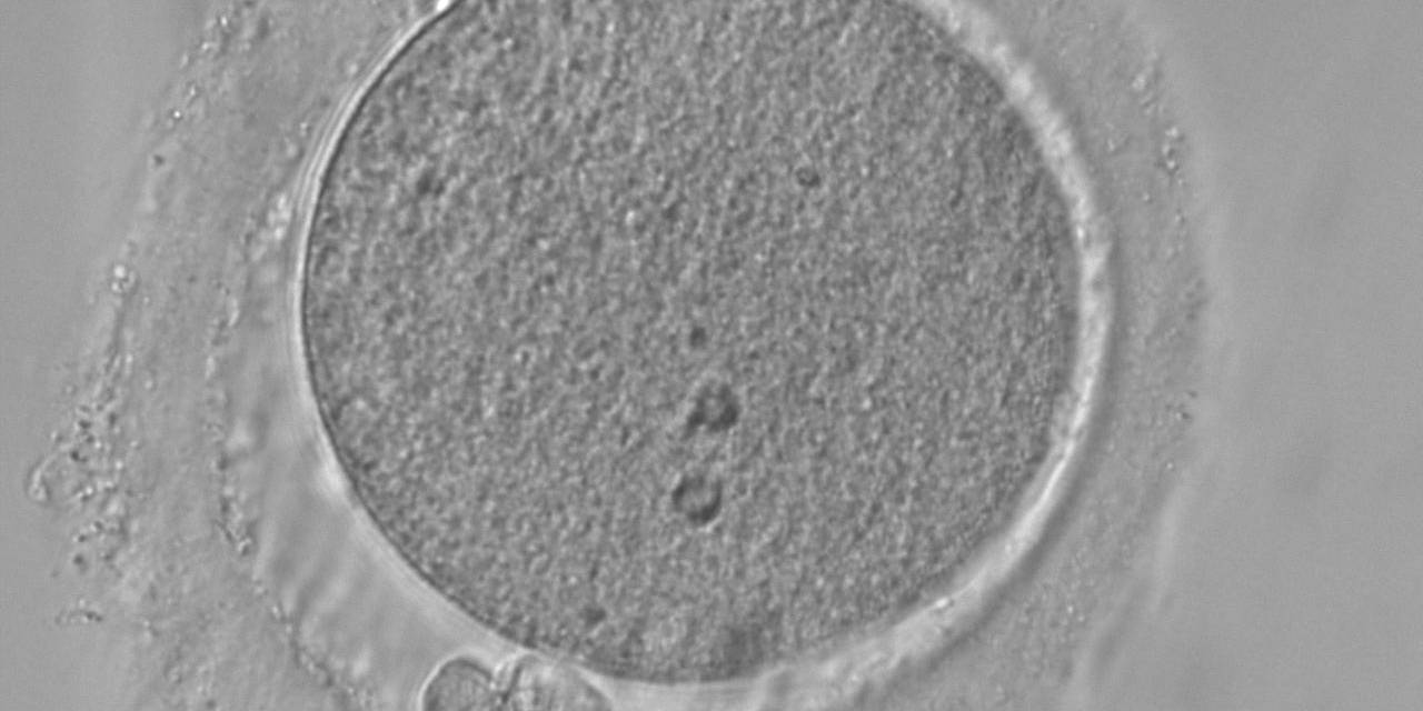
Figure 80
MII oocyte with a fragmented PBI (two pieces).
Sometimes it is difficult to distinguish between heavily fragmented PBI's and debris within the PVS (Figs 79–82). Fertilization and cleavage rate as well as embryo quality have been found to be unaffected by the presence of coarse granules in the PVS (Figs 83 and 84); however, the rates of implantation and pregnancy seem to be influenced (Hassan-Ali et al., 1998; Farhi et al., 2002). Granularity in the PVS has been associated with over-maturity of oocytes (Miao et al., 2009).
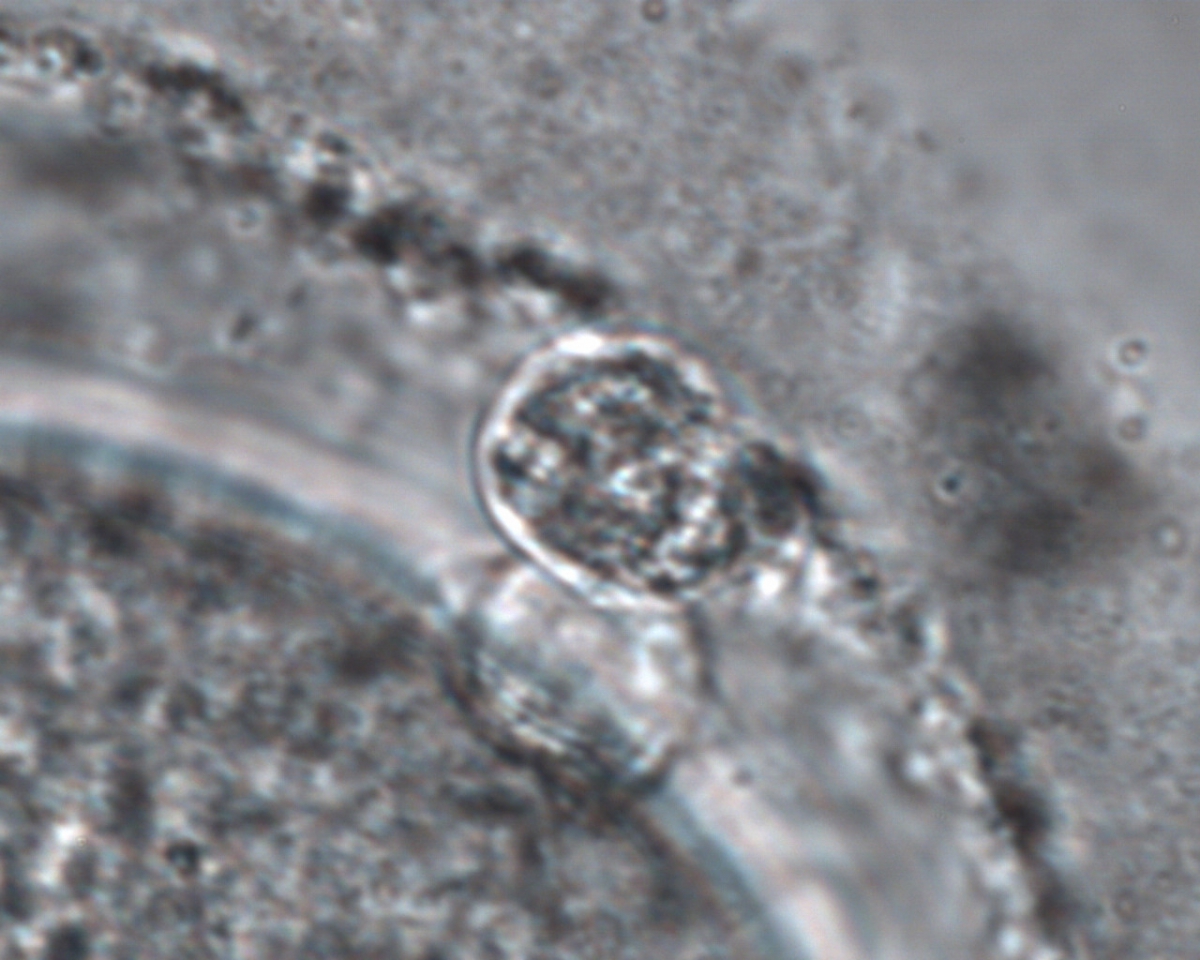
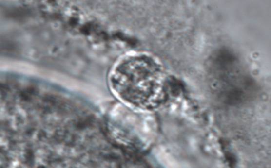
Figure 81
MII oocyte with a fragmented PBI (two pieces) (magnification ×1000).
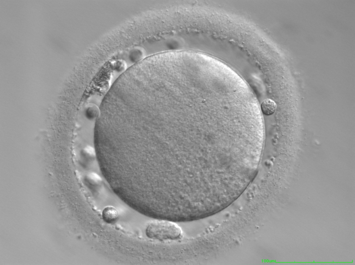
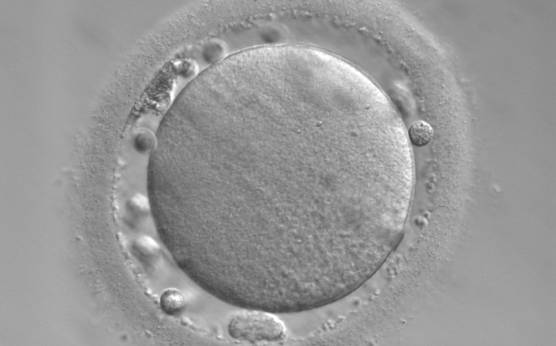
Figure 82
MII oocyte with a fragmented PBI and several cellular fragments within the PVS which are almost indistinguishable from PB1.
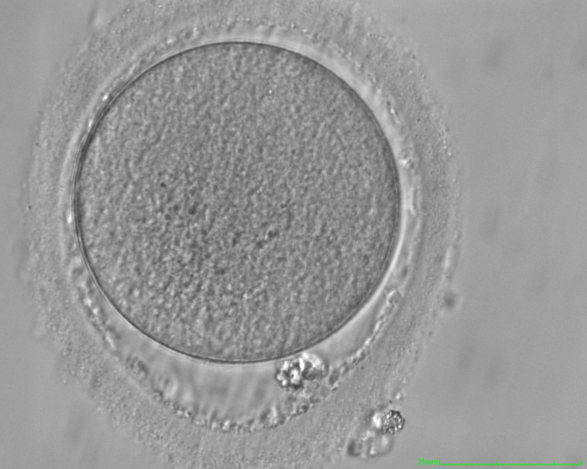

Figure 83
MII oocyte with a lobular/fragmented PBI and significant granulation in the The ZP has a dishomogeneous thickness and an irregular inner layer.
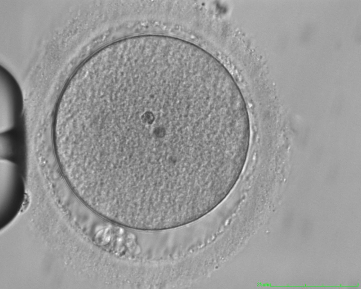
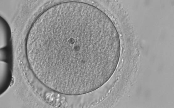
Figure 84
MII oocyte with a lobular/fragmented PBI and significant granulation in the PS. Two inclusion bodies can clearly be seen in the centre of the oocyte.
Article references:
Ciotti PM, Notarangelo L, Morselli-Labate AM, Felletti V, Porcu E, Venturoli S. First polar body morphology before ICSI is not related to embryo quality or pregnancy rate. Hum Reprod 2004;19:2334-2339.
Abstract/FREE Full Text
De Santis L, Cino I, Rabellotti R, Calzi F, Persico P, Borini A, Coticchio G. Polar body morphology and spindle imaging as predictors of oocyte quality. Reprod BioMed Online 2005;11:36-42.
CrossRef | Medline | Web of Science | Google Scholar
Ebner T, Moser M, Yaman C, Feichtinger O, Hartl J, Tews G. Elective transfer of embryos selected on the basis of first polar body morphology is associated with increased rates of implantation and pregnancy. Fertil Steril 1999;72:599-603.
CrossRef | Medline | Web of Science | Google Scholar
Ebner T, Moser M, Sommergruber M, Yaman C, Pfleger U, Tews G. First polar body morphology and blastocyst formation rate in ICSI patients. Hum Reprod 2002;17:2415-2418.
Abstract/FREE Full Text
Ebner T, Balaban B, Moser M, Shebl O, Urman B, Ata B, Tews G. Automatic user-independent zona pellucida imaging at the oocyte stage allows for the prediction of preimplantation development. Fertil Steril 2010;94:913-920.
CrossRef | Medline | Web of Science | Google Scholar
Esfandiari N, Burjaq H, Gotlieb L, Casper RF. Brown oocytes: implications for assisted reproductive technology. Fertil Steril 2006;86:1522-1525.
CrossRef | Medline | Web of Science | Google Scholar
Farhi J, Nahum H, Weissman A, Zahalka N, Glezerman M, Levran D. Coarse granulation in the perivitelline space and IVF-ICSI outcome. J Assist Reprod Genetics 2002;19:545-549.
CrossRef | Medline | Web of Science | Google Scholar
Fancsovits P, Tothne Z, Murber A, Takacs FZ, Papp Z, Urbancsek J. Correlation between first polar body morphology and further embryo development. Acta Biol Hung 2006;57:331-338.
CrossRef | Medline | Web of Science | Google Scholar
Hassan-Ali H, Hisham-Saleh A, El-Gezeiry D, Baghdady I, Ismaeil I, Mandelbaum J. Perivitelline space granularity: a sign of human menopausal gonadotrophin overdose in intracytoplasmic sperm injection. Hum Reprod 1998;13:3425-3430.
Abstract/FREE Full Text
Miao YL, Kikuchi K, Sun QY, Schatten H. Oocyte aging: cellular and molecular changes, developmental potential and reversal possibility. Hum Reprod Update 2009;15:573-585.
Abstract/FREE Full Text
Mikkelsen AL, Lindenberg S. Morphology of in-vitro matured oocytes: impact on fertility potential and embryo quality. Hum Reprod 2001;16:1714-1718.
Abstract/FREE Full Text
Montag M, Schimming T, Köster M, Zhou C, Dorn C, Rösing B, van der Ven H, Ven der Ven K. Oocyte zona birefringence intensity is associated with embryonic implantation potential in ICSI cycles. Reprod Biomed Online 2008;16:239-244.
Medline | Web of Science | Google Scholar
Shen Y, Stalf T, Mehnert C, Eichenlaub-Ritter U, Tinneberg HR. High magnitude of light retardation by the zona pellucida is associated with conception cycles. Hum Reprod 2005;20:1596-1606.
Abstract/FREE Full Text
Stanger JD, Stevenson K, Lakmaker A, Woolcott R. Pregnancy following fertilization of zona-free, coronal cell intact human ova. Hum Reprod 2001;16:164-167.
Abstract/FREE Full Text
Pelletier C, Keefe DL, Trimarchi JR. Noninvasive polarized light microscopy quantitatively distinguishes the multilaminar structure of the zona pellucida of living human eggs and embryos. Fertil Steril 2004;81:850-856.
CrossRef | Medline | Web of Science | Google Scholar
Rienzi L, Ubaldi FM, Iacobelli M, Minasi MG, Romano S, Ferrero S, Sapienza F, Baroni E, Litwicka K, Greco E. Significance of metaphase II human oocyte morphology on ICSI outcome. Fertil Steril 2008;90:1692-1700.
CrossRef | Medline | Web of Science | Google Scholar
Verlhac MH, Lefebvre C, Guillaud P, Rassinier P, Maro B. Assymetric division in mouse oocytes: with or without MOS. Curr Biol 2000;10:1303-1306.
CrossRef | Medline | Web of Science | Google Scholar
Xia P. Intracytoplasmic sperm injection: correlation of oocyte grade based on polar body, perivitelline space and cytoplasmic inclusions with fertilization rate and embryo quality. Hum Reprod 1997;12:1750-1755.
Abstract/FREE Full Text
