E. Cytoplasmic morphology assessment
E.1 Normal/granular
Homogeneous cytoplasm is expected in zygotes as for oocytes (Figs 190 and 191), but heterogeneous cytoplasm is of unknown developmental significance. Therefore, although some studies have reported that severe cytoplasmic anomalies in the zygote adversely affect the developmental and implantation potential of the resulting embryo (Kahraman et al., 2000; Ebner et al., 2003; Balaban and Urman, 2006), there is no clear evidence supporting these findings. Similarly, the presence of a peripheral cytoplasmic translucency in the fertilized oocyte (known as the ‘halo’; Fig. 192) or of minor dysmorphisms such as debris in the PVS (Fig. 193) or presence of refractile bodies in the cytoplasm (Fig. 194) have not been proved to be of prognostic value for implantation. Nevertheless, recording of these observations should be made as the accumulation of data could reveal some relevant links to developmental or implantation potential.
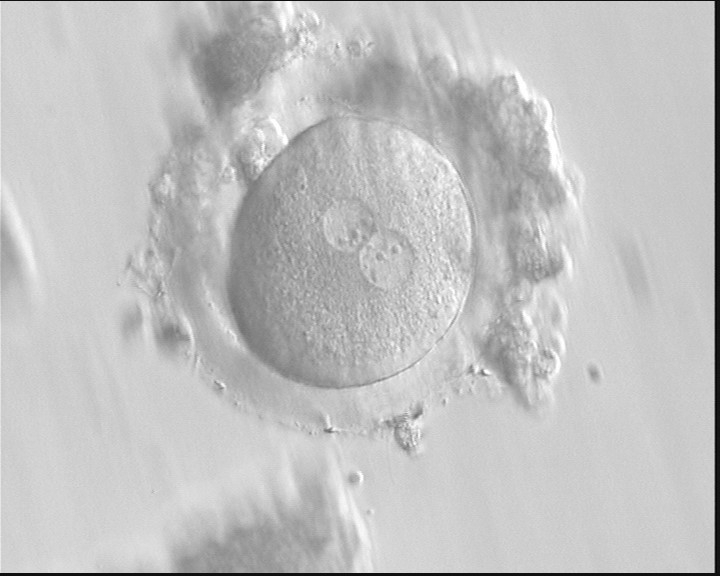
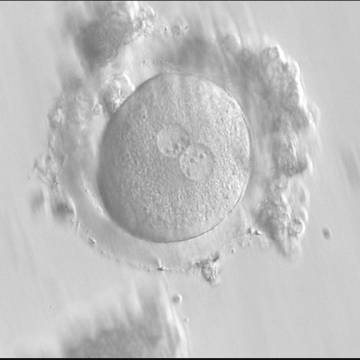
Figure 190
A zygote generated by IVF with frozen/thawed ejaculated sperm and observed at 16 h post-insemination showing normal cytoplasmic morphology (400× magnification). NPBs are obviously different in the two PNs. It was transferred but failed to implant.
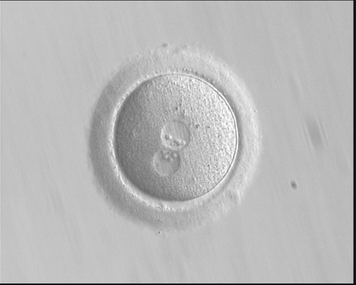
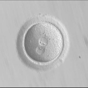
Figure 191
A zygote generated by ICSI, which subsequently underwent polar body biopsy, shows normal cytoplasmic morphology (400× magnification). It was discarded due to aneuploidy.
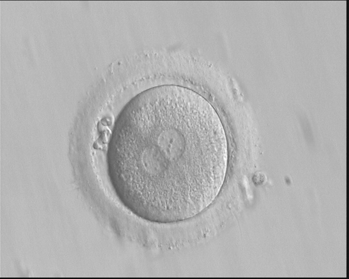
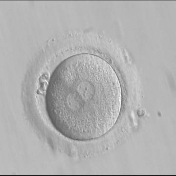
Figure 192
A zygote generated by IVF with frozen/thawed ejaculated sperm and observed at 16 h post-insemination showing normal cytoplasmic morphology with an evident clear cortical zone, the halo, in the cytoplasm (400× magnification). NPBs are scattered and different-sized, polar bodies are fragmented. It was cryopreserved.
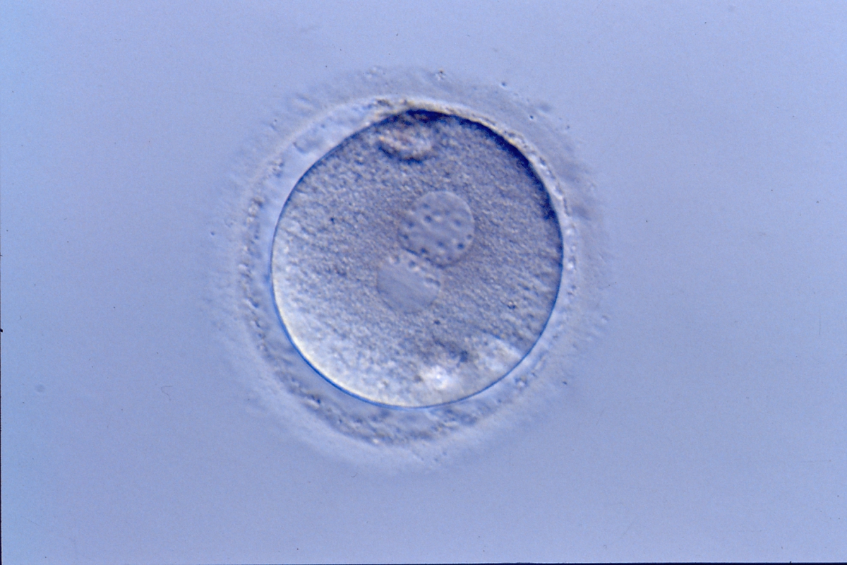
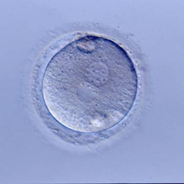
Figure 193
A zygote generated by ICSI showing normal cytoplasmic morphology, a thin ZP and small debris in an enlarged PVS (400× magnification). It was transferred but clinical outcome is unknown.

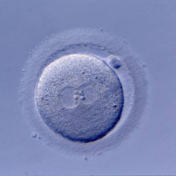
Figure 194
A zygote generated by ICSI showing normal cytoplasmic morphology except for a refractile body visible at the 3 o'clock position in this view (400× magnification). It was transferred but clinical outcome is unknown.
Normal cytoplasm is clearly distinguishable from granular cytoplasm, but to make comparative observations attention should be paid to the optical system and culture medium employed. The severity of granularity is generally based on the diameter and depth of the granular area that may occupy either the whole zygote (Fig. 195), or small (Figs 196–198) or large areas of the cytoplasm (Figs 199 and 200).


Figure 195
A zygote generated by ICSI displaying heterogeneous, granular cytoplasm (200× magnification). NPBs are large-sized and polar bodies are fragmented. It was discarded due to poor subsequent development.
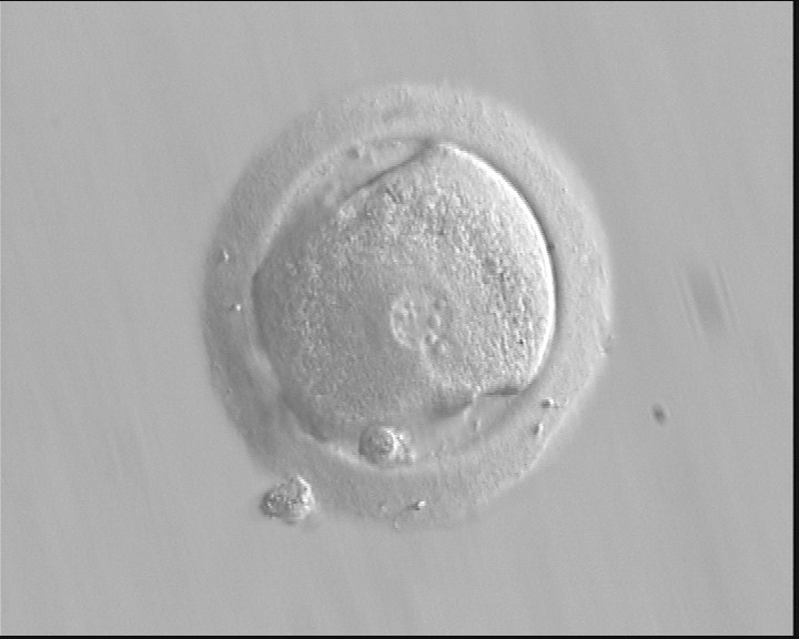
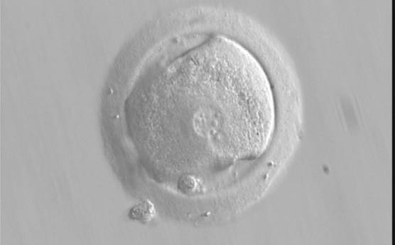
Figure 196
A zygote generated by IVF using frozen/thawed ejaculated sperm and observed at 17 h post-insemination showing an irregular oolemma and dysmorphic granular cytoplasm (400× magnification). PNs are different in size and peripherally located. NPBs differ in size and number between PNs. It was transferred but failed to implant.
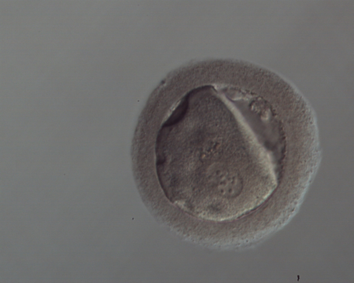
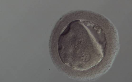
Figure 197
A zygote generated by ICSI with peripheral PNs (150× magnification). The oolemma is irregular and the cytoplasm is dysmorphic and granular. The ZP is thick and dark. It was discarded.
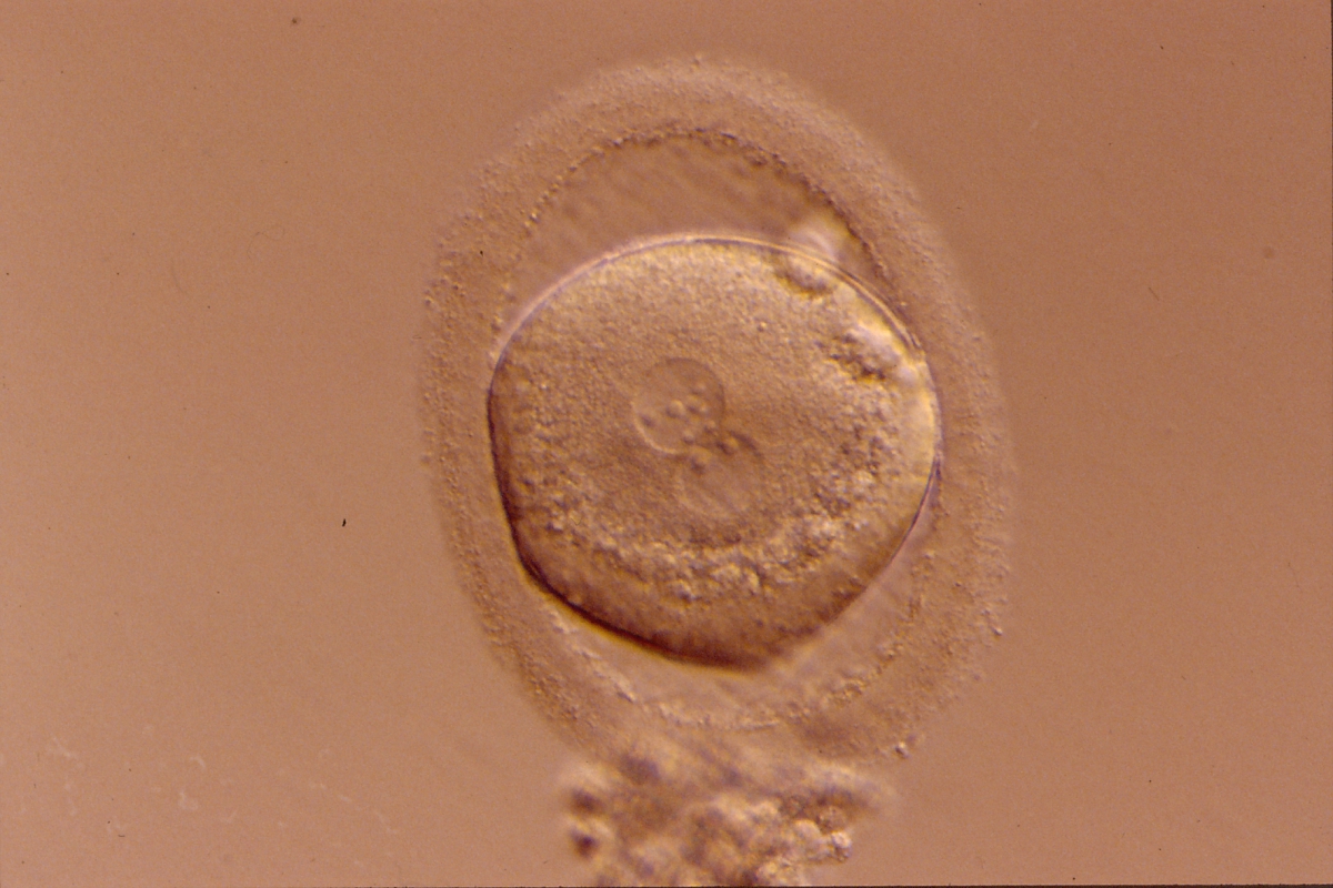
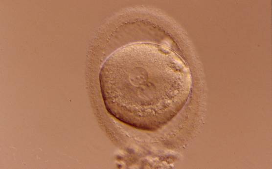
Figure 198
A zygote generated by ICSI displaying granular cytoplasm, especially in the area immediately adjacent to the clear cortical zone (400× magnification). There is an enlarged PVS and an ovoid ZP. NPBs differ in number and size. It was transferred but clinical outcome is unknown.

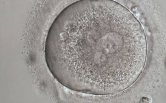
Figure 199
A zygote generated by ICSI with four PNs (possibly a result of fragmentation of an originally normal-sized PN), displaying very granular cytoplasm and a clear cortical zone (600× magnification). It was discarded.

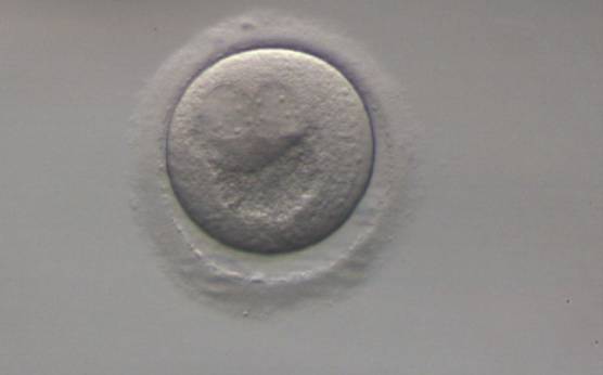
Figure 200
A zygote generated by ICSI using frozen/thawed ejaculated sperm (200× magnification). PNs are peripherally located with a large inclusion positioned directly below the PNs that is displaying a crater-like appearance as a consequence of severe organelle clustering. It was discarded.
It has been reported that half of the oocytes with dysmorphic phenotypes such as organelle clustering are aneuploid, with hypohaploidy being the predominant abnormality (Van Blerkom and Henry, 1992). This severe cytoplasmic disorganization is associated with a lower intracytoplasmic pH and decreased ATP content (Van Blerkom et al., 1997). These dysmorphic changes would be inherited in the zygote. Apparently, intracytoplasmic organelle clustering (Fig. 200) is a type of severe abnormality that is significantly repetitive in consecutive cycles and is a negative predictor of pregnancy and implantation rates, although the cleavage stage embryo quality, as observed by light microscopy, is apparently not affected (Meriano et al., 2001).
E.2 Small vacuoles/large vacuoles
The presence of a few small vacuoles (diameter of 5–10 μm) that are apparently fluid filled and transparent (Figs 201 and 202) have not been associated with detectable biological consequences, but some concern may arise when several vacuoles appear (Figs 203 and 204) or appear with other morphological anomalies (Fig. 205).
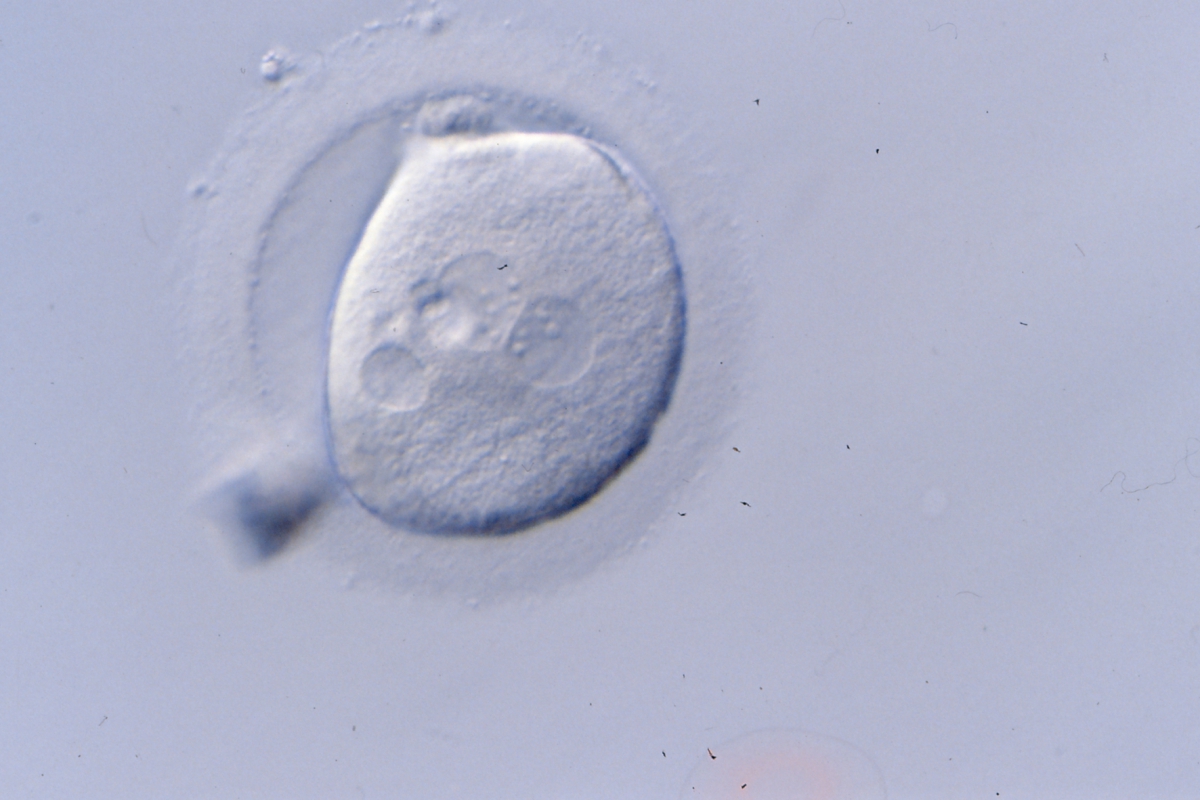

Figure 201
An irregularly shaped zygote generated by ICSI showing centrally positioned PNs and a medium-sized vacuole at the 8 o'clock position in the cytoplasm (400× magnification). It was transferred but clinical outcome is unknown.
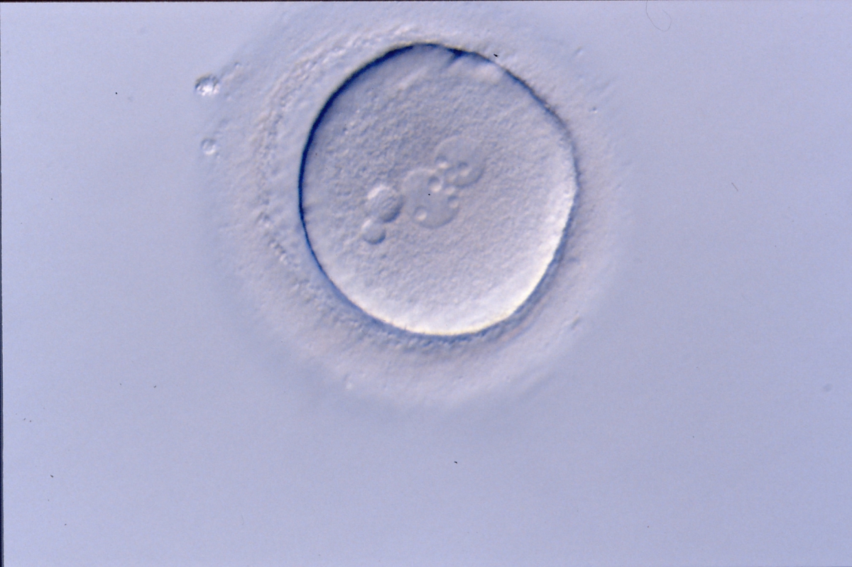
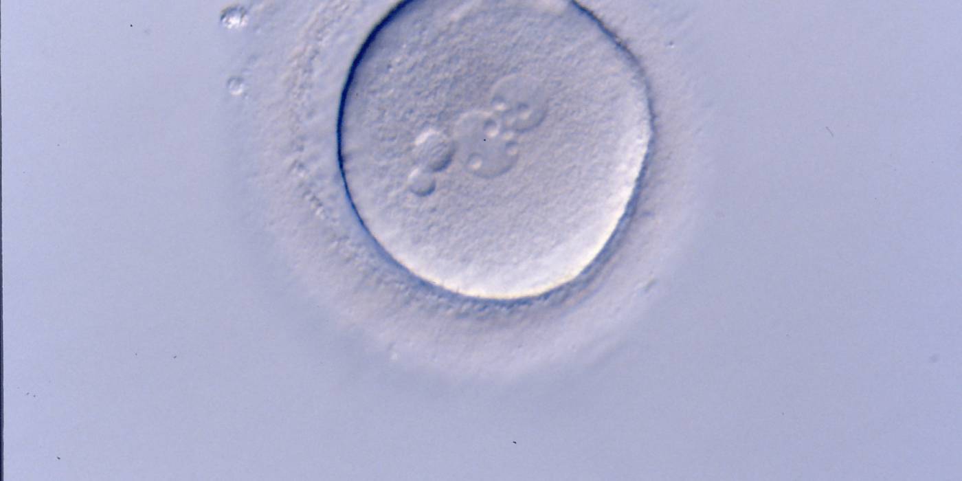
Figure 202
A zygote generated by ICSI with centrally positioned different-sized PNs, displaying two small vacuoles at the 9 o'clock position in the cytoplasm (400× magnification). It was transferred but clinical outcome is unknown.
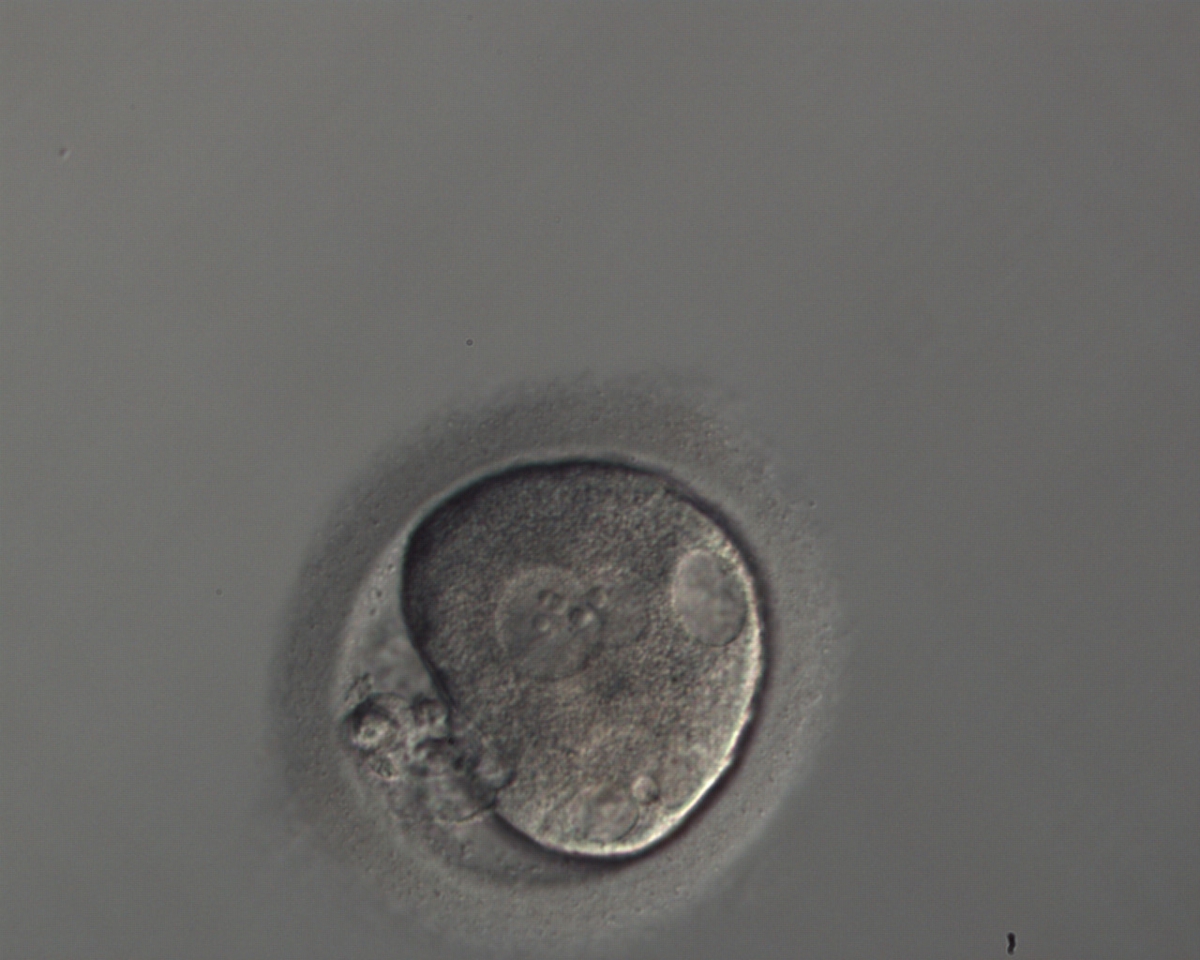
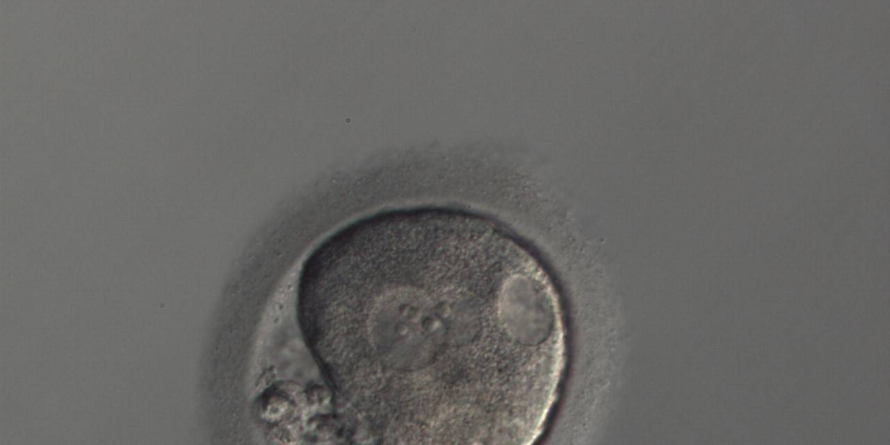
Figure 203
A zygote generated by ICSI with an irregular oolemma and a large PVS with possibly highly fragmented polar bodies (150× magnification). Different sized overlapping PNs are peripherally positioned and several vacuoles of different sizes are present at the 2, 5 and 6 o'clock position in the cytoplasm. It was discarded.
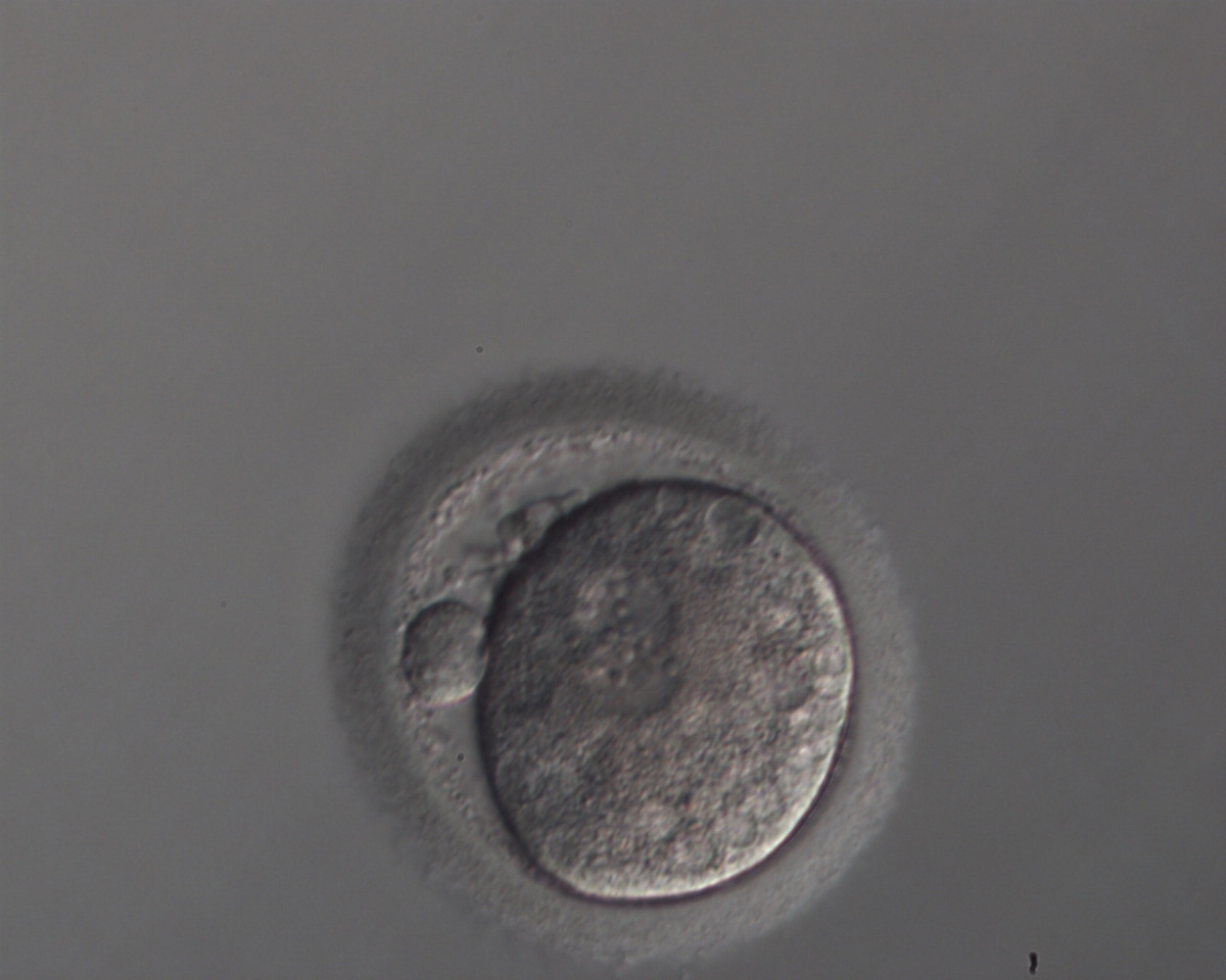
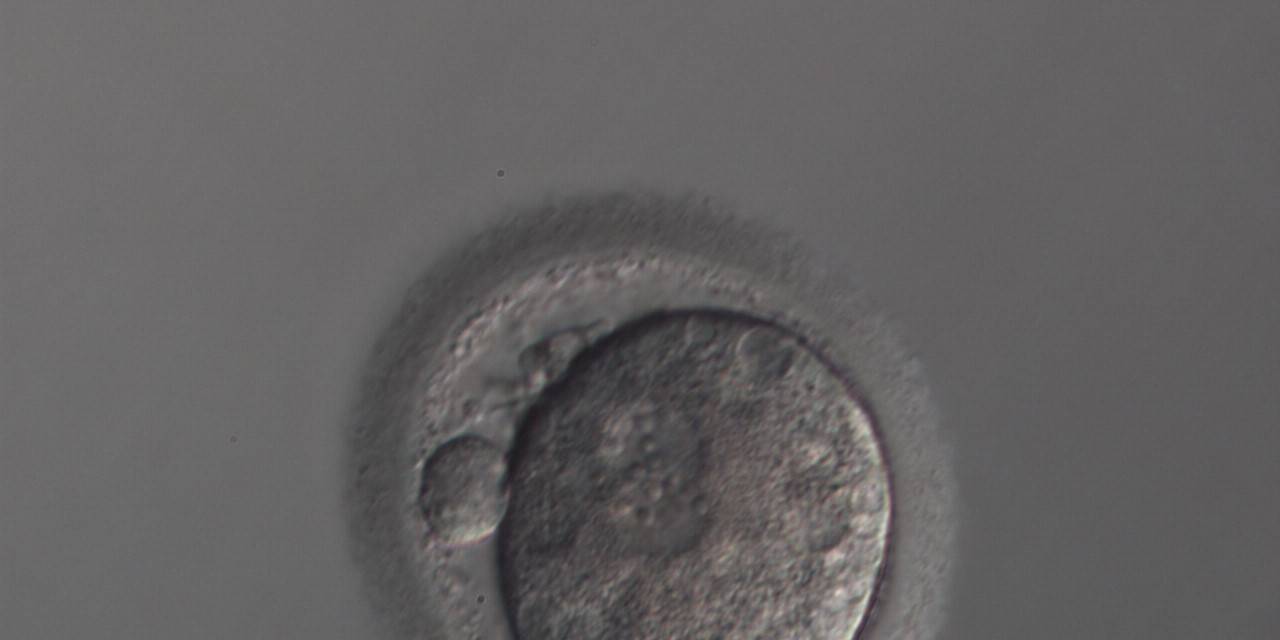
Figure 204
A zygote generated by ICSI with slightly overlapping peripherally positioned PNs and a large PVS with one large and one fragmented polar body (150× magnification). Many small vacuoles are present throughout the cytoplasm. It was discarded.

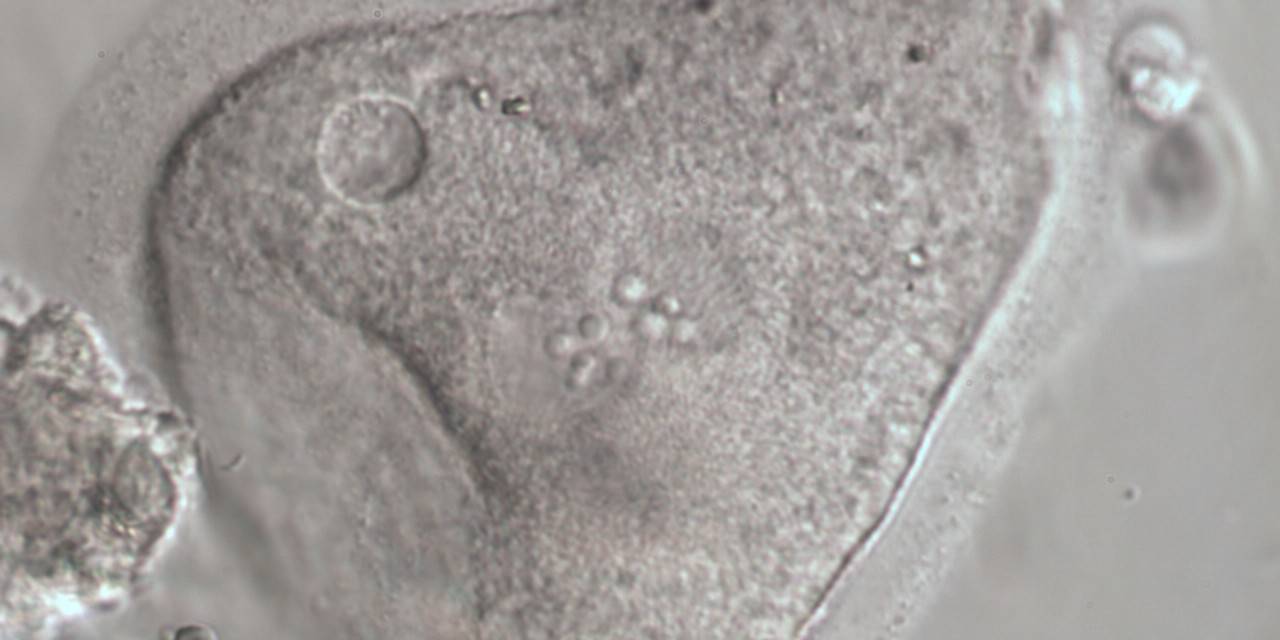
Figure 205
Severely dysmorphic zygote generated by IVF showing small PNs and an irregularly shaped ZP and oolemma and lack of a PVS (600× magnification). There is a small vacuole present at 10 o'clock with refractile bodies immediately adjacent. It was discarded.
Large vacuoles (>14 μm in diameter) in fertilized oocytes (Figs 206–210) can interfere with cleavage planes, resulting in reduced blastocyst formation (Ebner et al., 2005). For this reason, they are normally not considered for transfer.

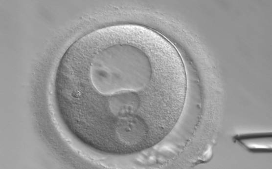
Figure 206
A zygote generated by ICSI showing peripherally positioned PNs and a very large vacuole at the 12 o′clock position in the cytoplasm (400× magnification). Polar bodies are fragmented and the ZP is of irregular thickness. It was discarded.
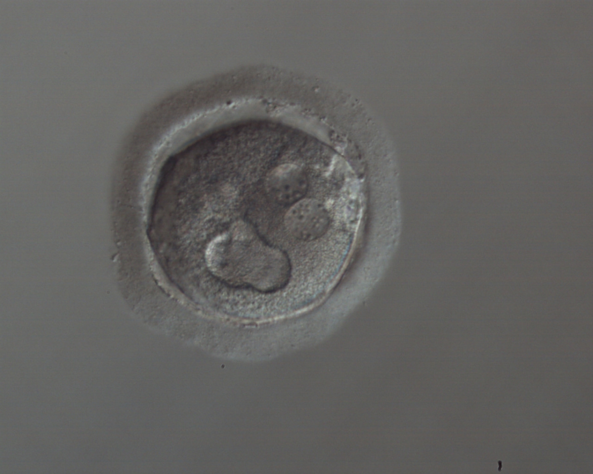
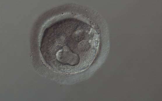
Figure 207
A zygote generated by ICSI showing cytoplasmic abnormalities (150× magnification). The PNs are juxtaposed and peripherally positioned with a large vacuole of irregular shape at the 6 o'clock position in the cytoplasm. The cytoplasm is granular and the ZP is thick and heterogeneous in appearance. It was discarded.
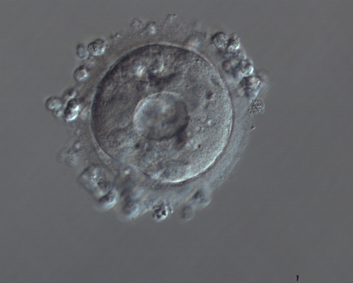
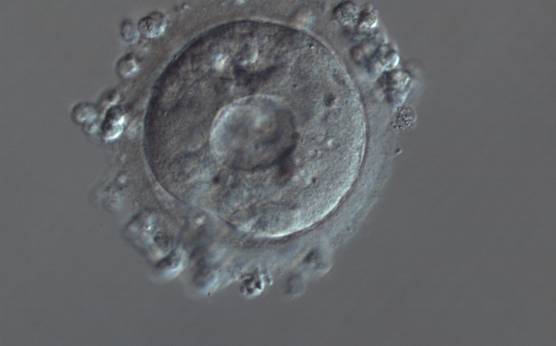
Figure 208
A zygote generated by ICSI showing severe cytoplasmic abnormalities (150× magnification). There is a large centralized vacuole with many small vacuoles surrounding it in a granular cytoplasm. It was discarded.
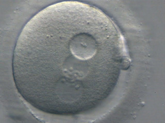
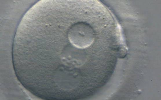
Figure 209
A zygote generated by ICSI showing peripherally located PNs with the same number and size of NPBs perfectly aligned at the PN junction (400× magnification). There is a large vacuole immediately adjacent to the two PNs that is almost the same size as the PNs. It was discarded.
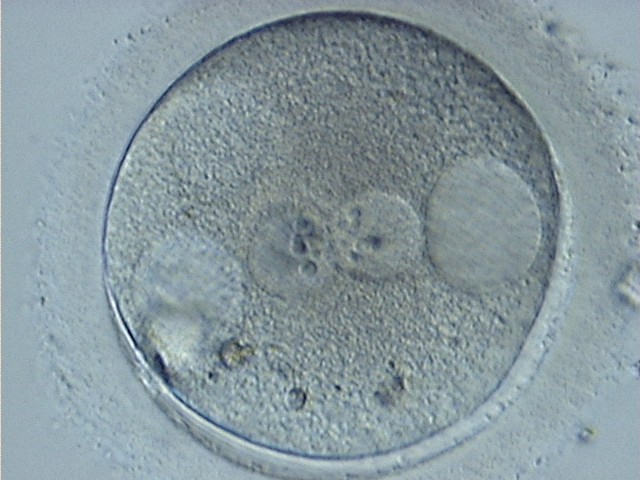
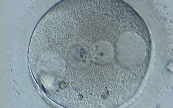
Figure 210
A zygote generated by ICSI showing centrally positioned PNs and two large vacuoles immediately adjacent to each of the PNs at the 3 and 9 o'clock positions in the cytoplasm (400× magnification). There are also refractile bodies present at the 6–7 o'clock positions and an area of clustering at 11 o'clock. It was discarded.
At the time of fertilization check and especially after conventional IVF, it is extremely important to carefully score the cytoplasm for the presence of SER discs (see Chapter One), which are associated with the risk of a deleterious clinical outcome (Otsuki et al., 2004).
Article references:
Balaban B, Urman B. Effect of oocyte morphology on embryo development and implantation. Reprod BioMed Online 2006;12:608-615.
Medline | Web of Science | Google Scholar
Kahraman S, Yakin K, Dönmez E, Samli H, Bahçe M, Cengiz G, Sertyel S, Samli M, Imirzalioğlu N. Relationship between granular cytoplasm of oocytes and pregnancy outcome following intracytoplasmic sperm injection. Hum Reprod 2000;15:2390-2393.
Abstract/FREE Full Text
Ebner T, Moser M, Sommergruber M, Gaiswinkler U, Wiesinger R, Puchner M, Tews G. Presence, but not type or degree of extension, of a cytoplasmic halo has a significant influence on preimplantation development and implantation behaviour. Hum Reprod 2003;18:2406-2412.
Abstract/FREE Full Text
Ebner T, Moster M, Sommergruber M, Gaiswinkler U, Shebl O, Jesacher K, Tews G. Occurrence and developmental consequences of vacuoles throughout preimplantation development. Fertil Steril 2005;83:1635-1640.
CrossRef | Medline | Web of Science | Google Scholar
Meriano JS, Alexis J, Visram-Zaver S, Cruz M, Casper RF. Tracking of oocyte dysmorphisms for ICSI patients may prove relevant to the outcome in subsequent patient cycles. Hum Reprod 2001;16:2118-2123.
Abstract/FREE Full Text
Otsuki J, Okada A, Morimoto K, Nagai Y, Kubo H. The relationship between pregnancy outcome and smooth endoplasmic reticulum clusters in MII human oocytes. Hum Reprod 2004;19:1591-1597.
Abstract/FREE Full Text
Van Blerkom J, Henry G. Oocyte dysmorphism and aneuploidy in meiotically mature human oocytes after ovarian stimulation. Hum Reprod 1992;7:379-390.
Abstract/FREE Full Text
Van Blerkom J, Antczak M, Schrader R. The developmental potential of the human oocyte is related to the dissolved oxygen content of follicular fluid: association with vascular endothelial growth factor levels and perifollicular blood flow characteristics. Hum Reprod 1997;12:1047-1055.
Abstract/FREE Full Text
